thiamine deficiency - Page 4

Unraveling Symptoms and Syndromes

15252 views

What Is a Syndrome?

A syndrome is the name given to a collection of symptoms and physical signs that have been observed in the past in a single patient or in a group of similar patients. This is often named after the first person to report this set of observations. It is called a syndrome when others have made the same observations, sometimes years later. The terminology is purely descriptive, even though there may be a constellation of abnormal laboratory tests associated with the clinical facts. Unfortunately, the underlying cause is seldom, if ever, known.

Chronic Fatigue Syndrome

Chronic fatigue syndrome (CFS) is also known as myalgic encephalomyelitis (ME). In a review, it is described as a “challenge to physicians”. Its prevalence is reported as approximately 1% in the general German population. The author states that there are no convincing models that might explain the underlying cause as an independent unique disease. A variety of conditions such as chronic infectious disease, multiple sclerosis, endocrine disorders and psychosomatic disease are suggested in a differential diagnosis. There is said to be a significant overlap with major depression.

Another review describes CFS as characterized by debilitating fatigue that is not relieved with rest and is associated with physical symptoms. In order to make the diagnosis, these authors indicate that at least four of the following symptoms are required to make the diagnosis. They include feeling unwell after exertion, unrefreshing sleep, impaired memory or concentration, muscle pain, aching joints, sore throat, or new headaches. They also say that no pharmacologic or alternative medicine therapies have been proven effective.

Fibromyalgia Syndrome

According to the American College of Rheumatology, fibromyalgia syndrome (FMS) is a common health problem characterized by widespread pain and tenderness. Although chronic, there is a tendency for the pain to fluctuate in intensity and location around the body. Deficient understanding of its true cause gives rise to the false concept that it is neurotic. It is associated with chronic fatigue and patients often have sleep disorders. It is estimated that it affects 2 to 4% of the general population and is most common in women. It affects all ages and the causes are said to be unclear. FMS patients may require psychiatric therapy due to accompanying mental problems. Gonzalez and associates concluded that the combination of psychopathological negative emotionality, interpersonal isolation and low hedonic capacity that they found in a group of patients has implications for the daily living and treatment of these patients.

Regional Pain Syndrome

Complex regional pain syndrome (CRPS) is another common and disabling disorder, characterized by defective autonomic nervous system function and inflammatory features. It reportedly occurs acutely in about 7% of patients who have limb fractures, limb surgery, or other injuries, often quite minor. A small subset of patients progress to a chronic form in which autonomic features dominate. Allodynia (pain due to a stimulus that does not usually provoke pain) and hyperalgesia (increased pain from a stimulus that usually provokes pain) are features of CRPS and require a better understanding.

Sleep Apnea Syndrome

Apnea is the term used for a temporary cessation of automatic breathing that usually happens during the night. This syndrome is described as the most common organic disorder of excessive daytime somnolence. Its prevalence is highest among men age 40 to 65 years and may be as high as 8.5% or higher in this population. It is associated with cigarette smoking, use of alcohol and poor physical fitness.

Similar Cause with Different Manifestations

Complex Regional Pain Syndrome is related to microcirculation impairment associated with tissue hypoxia (lack of oxygen) in the affected limb. Without going into the complex details, hypoxia induces a genetic mechanism called hypoxia inducible factor (HIF-1 alpha) that has a causative association with CRPS. It has been found that inhibiting this factor produced an analgesic effect in a mouse model. The interesting thing about this is that thiamine deficiency does exactly the same thing because it induces biochemical effects similar to those produced by hypoxia (pseudo[false]hypoxia). A group of physicians in Italy have shown that high doses of thiamine produced an appreciable improvement in the symptoms of three female patients affected by fibromyalgia and are probably pursuing this research. Dietary interventions have been reported in seven clinical trials in which five reported improvement. There was variable improvement in associated fatigue, sleep quality, depression, anxiety and gastrointestinal symptoms.

Dr. Marrs and I have published a book that emphasizes deficient energy metabolism as a single cause of many, if not all, diseases. The symptomatic overlap in these so-called syndromes is generated by defective function of cellular metabolism in brain. Fatigue is the best symbol of energy deficiency and the English translation of the Chinese word beriberi is “I can’t, I can’t”. Fatigue is a leading symptom in beriberi. When physicians diagnose psychosomatic disease as “it’s all in your head”, they are of course, quite right. However, to imagine or conclude that the variable symptoms that accompany the leading one of fatigue are “imaginary” is practically an accusation of malingering. The brain is trying to tell its owner that it has not got the energy to perform normally and the physician should be able to recognize the problem by understanding the mechanism by which the symptoms are produced. Every thought, every emotion, every physical action, however small, requires the consumption of energy. Obviously we are considering variable degrees of deficiency from slight to moderate. The greater the deficiency the more serious is the manifestation of disease that follows. Death is a manifestation of deficiency that no longer permits life.

Our book is written primarily for physicians, but it is sufficiently lacking in technological language to encourage reading by patients. It emphasizes, by descriptions of case after case, the details of how genetic risk and failed brain energy are together unable to meet and adapt a person’s ability to meet the daily stresses of life. Stress, genetic risk and poor diet all go together. A whole chapter discusses the functions of the autonomic nervous system and how it deviates when the control mechanisms in the lower brain are defective. This system is the nervous channel that enables the brain to communicate the adaptive body actions necessary to meet living in an essentially hostile environment. We show that an excess of sugar and/or alcohol produce deficiency of vitamin B1 and the so-called psychosomatic disease that results is really early beriberi “I can’t, I can’t”. Variability in symptoms caused by this effect is because the cellular energy deficiency distribution varies from person to person and is affected by genetically determined differences.

This is illustrated by the case of a boy with eosinophilic esophagitis whose first eight years of life were marked by repeated diagnoses of psychosomatic disease. At the age of eight, upper endoscopy revealed the pathology in the esophagus. There was a family history of alcoholism and he was severely addicted to sugar. Many of his symptoms cleared with the administration of a thiamine derivative and resulted in a dramatic increase in stature. No pediatrician or other physician whose attendance was sought through those first 8 years evidently had ever questioned diet or the gross ingestion of sweets. They simply treated each condition as a confirmation that they were “psychological”.

It is worth noting that references 1 through 4 refer to both CFS and FMS syndromes being affected by psychological issues. This implies that the patient is “inventing” the poorly understood (and often bizarre) symptoms as a result of neurosis. The unfortunate complainant may easily become classified in the mind of the attendant physician as a “problem patient”. I have become aware that this can rise to such a degree of misunderstanding that the patient is denied access to the physician and even to other physicians in the same clinic. It is indeed about time that an overall revision be made to the absurd concept that the brain can “invent” a sensation that has no importance in solving the electrochemical problem. When we see the statistics of incidence of these common syndromes we have to conclude that there is an underlying cause and effect that pervades the general population. We are very conscious that our cars need the right fuel to work efficiently but rarely take it into consideration that the quality of food is our sole source of energy synthesis.

We Need Your Help

More people than ever are reading Hormones Matter, a testament to the need for independent voices in health and medicine. We are not funded and accept limited advertising. Unlike many health sites, we don’t force you to purchase a subscription. We believe health information should be open to all. If you read Hormones Matter, like it, please help support it. Contribute now.

Yes, I would like to support Hormones Matter. 

Image created using Canva AI.

This article was published originally December 2019. 

Rest in peace Dr. Lonsdale. May 2024.

Thiamine Insufficiency Relative to Carbohydrate Consumption

30490 views

Thiamine (vitamin B1) is an essential micronutrient responsible for key reactions involved in the conversion of the foods we consume into the chemical energy substrate requisite for cellular function, adenosine triphosphate (ATP). Absent sufficient ATP, all sorts of metabolic functions become disordered leading to the disease processes that dominate western medicine. Chronic inflammation, altered immune function, hormone dysregulation, cognitive and mood disorders, and dysautonomias, all can be traced back to insufficient thiamine > inefficient mitochondrial function, reduced ATP, and the compensatory reactions that ensue.

Among the most common but least well-recognized contributors to thiamine deficiency is the regular consumption of a high carbohydrate/highly processed food diet. Although most of these foods are enriched or fortified with thiamine, perhaps staving off more severe deficiencies, the density of sugars overwhelms mitochondrial capacity to process these foods, both the thiamine and any other potential nutrients are excreted, while the carbohydrates themselves are stored as fat for future use. High-calorie malnutrition is a common contributor to thiamine deficiency in obesity but also may develop in presumed healthy athletes whose diets focus heavily on high carbohydrate intake.

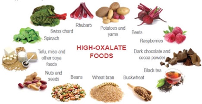
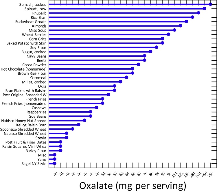
Thiamine, along with other B vitamins is often deficient in vegetarian and vegan diets as well. Not only do fruits, vegetables, and carbohydrates contain minimal, if any, thiamine, but some have anti-thiamine factors and are high in what are called oxalates. Anti-thiamine factors found in some fruits and vegetables interfere with the absorption or digestion of thiamine. Oxalates are mineralized crystals of sorts that tend to build up and store in places like the kidneys (kidney stones), but also may store and cause problems anywhere in the body like bones, arteries, eyes, heart, and nerves. Effective oxalate metabolism and clearance requires thiamine. Since vegetarian and vegan diets are also carbohydrate intensive, thiamine deficiency and oxalate issues may be compounded. Thus, a number of common diets not only contain reduced thiamine content but cause an increased need for thiamine by at least three mechanisms; higher carbohydrate consumption overwhelming capacity, which is then magnified by poor carbohydrate and oxalate processing.

Add daily coffee, tea, and/or alcohol consumption to any diet, and whatever thiamine that is consumed is either inactivated by enzymes before being used or is unabsorbable. Add a medication or four and thiamine availability will tank simultaneously with an increased need. Medications both block nutrient uptake and/or increase the need for nutrients by inducing mitochondrial damage. Given that 70% percent of the US population takes at least one medication regularly, while 20% take four or more, it is safe to say, that a good percentage of the population is consuming insufficient thiamine to maintain mitochondrial function and health.

Are We Really Thiamine Deficient?

As an essential nutrient, thiamine must be consumed regularly to maintain sufficient concentrations. The question is how much thiamine is sufficient to maintain health? Current RDA values for daily thiamine intake suggest a little over a milligram per day is adequate for most adults. If this is true, then the minimum value can be attained through just about any diet including those dominant in highly processed, carbohydrate-dense foods, which are commonly either enriched or fortified with thiamine. Everything from bread to cereals and even junk food like Oreos have thiamine. Per the RDA values, none of us ought to be thiamine deficient and none of us ought to require thiamine supplementation, and yet, many of us are and do. Indeed, several studies, across disparate populations show that even by this minimum standard, deficiency is a serious health problem. From our book:

  • 76% of diabetics (type 1 and type 2)
  • 29% of obese patients, 49% of post-bariatric surgery
  • 40% of community-dwelling elderly, 48% of elderly patients in acute care
  • 55% of cancer patients
  • 20% ER patients (random sample, UK)
  • 33% of congestive heart failure patients
  • 38% of pregnant women, more with hyperemesis
  • 30% of psychiatric patients

It takes approximately 18 days to completely abolish endogenous thiamine stores in a diet that is completely devoid of thiamine. Except under total starvation, medical or industrial food production mishaps, and experimentally contrived situations, thiamine consumption is never completely abolished. It waxes and wanes by dietary choices and life stressors. According to rodent studies, it takes a reduction of greater than 80% of thiamine stores before the more severe neurological symptoms are recognizable. In humans, these symptoms include those associated with Wernicke’s encephalopathy, the various forms of beriberi, and dysautonomic function. These include but are not limited to: ataxia, changes in mental status, optic neuritis, ocular nerve abnormalities, diminished visual acuity, high-output cardiac failure with or without edema, high pulse pressure, polyneuropathy (sensorimotor), enteritis, esophagitis, gastroparesis, nausea and vomiting, constipation, hyper- or hypo-stomach acidity, sympathetic/parasympathetic imbalance, postural orthostatic tachycardia syndrome (POTS), cerebral salt wasting syndrome, vasomotor dysfunction, respiratory distress, reduced vital capacity, and/or low arterial O2, high venous O2.

With a less severe thiamine deficiency, symptoms are rarely recognized as such and often attributed to psychological manifestations. A not entirely ethical study done in 1942 involving 11 women on a low thiamine diet over a period of ~3-6.5 months found striking symptoms.

  • During this time all subjects showed definite changes in personality.
  • They became irritable, depressed, quarrelsome, and uncooperative.
  • Two threatened suicide. All became inefficient in their work, forgetful, and lost manual dexterity.
  • Their hands and feet frequently felt numb.
  • Headaches, backaches, sleeplessness, and sensitivity to noises were noted.
  • The subjects fatigued easily and were not able to vigorous exertion.
  • Constipation was the rule, but no impairment, of gastrointestinal motility, could be demonstrated fluoroscopically.
  • Anorexia, nausea, vomiting, and epigastric distress were frequently observed.
  • Low blood pressure and vasomotor instability were present in all patients.
  • At rest, pulse rates were low (55 to 60 per minute) but tachycardia followed moderate exertion. Sinus arrhythmia was marked.
  • Macrocytic, hypochromic anemia of moderate severity (3.0 to 3.5 million red cells) developed in 5 cases.
  • A decrease in serum protein concentration occurred in 8 subjects.
  • Basal metabolic rates were lowered by 10 to 33 points.
  • Fasting blood sugar was often abnormally high.

The study above demonstrated a rapid and dramatic onset of symptoms relative to a diet with limited thiamine. Depending upon caloric intake, the amount of thiamine allowed was approximately 1/3 to 1/5 of the amount recommended by the RDA. Admittedly, the RDA for thiamine is low, to begin with, but even so, this was not a complete absence of thiamine. Since the study took place in the early 1940s, it is difficult to ascertain the specifics of the diet. Nevertheless, it demonstrates a clear association between general health and one’s ability to function, and thiamine insufficiency.

High Carbohydrate Diets Equal Lower Thiamine

More recently, a short and very small study (12 days and 12 participants) of active young men and women (ages 25-30) investigated the relationship between carbohydrate intake and thiamine status. Thiamine was measured in blood, plasma, urine (creatinine), and feces at four time points: at baseline, before the study began, during an adaptation phase where carbohydrate intake represented 55% of the total caloric intake, and during the two subsequent intervention phases, where carbohydrate intake was increased to 65% and 75% of the total caloric intake, respectively. Both caloric and thiamine intake was held constant throughout the study despite the increased intake of carbohydrates. Activity levels were also held constant. Across this short-term study, as carbohydrate intake increased, plasma, and urinary thiamine decreased. Excretion through feces remained unchanged. Transketolase enzyme activity was also measured but remained unchanged. Given the short-term nature of this study, the fact that transketolase remained unchanged is unexpected. In addition to the decreasing thiamine values, there were several changes in lipid profile as well. Despite the short duration of this study, however, the results show a clear relationship between carbohydrate intake and thiamine status; one that would likely be magnified over time and certainly if other life stressors and medical and environmental toxicants were added to the mix.

It is important to note current dietary guidelines suggest carbohydrate consumption should fall between 45-65% of total calories, percentages which, per this study would decrease thiamine availability significantly. From the baseline diet to the 55% adaptation phase, thiamine dropped precipitously, only to drop even further at the 65% phase. A recent study surveying macronutrient consumption showed that average carbohydrate consumption across the US population represented approximately 50% of total caloric intake. Importantly though, the study found that 42% of the carbohydrate consumption came in the form of what researchers termed ‘low-quality carbs’ e.g. sugary processed foods with no nutritional value. Thiamine is only found in pork, beef, wheat germ and whole grains, organ meats, eggs, fish, legumes, and nuts. It is not present in fats/oils, polished rice, or simple sugars, nor are dairy products or many fruits and vegetables a good source. Indeed as mentioned previously, some fruits and vegetables may contain anti-thiamine factors. A diet that is 42% empty calories, that contains limited to no nutritive value, save except what has been added post hoc via enrichment, begs for mitochondrial damage and the illnesses that ensue. And yet, that is precisely the nutritional landscape in which most of us exist.

Admittedly, both studies were very small, but the research connecting thiamine deficiency to ill-health and carbohydrate consumption to thiamine loss is clear. Given the dominance of ultra-processed carbohydrate-dense foods in the modern diet, is likely that high-calorie malnutrition underlies much of the chronic illness that plagues western medicine. To learn more about thiamine deficiency and the havoc it wreaks on health: Thiamine Deficiency Disease, Dysautonomia, and High Calorie Malnutrition.

We Need Your Help

More people than ever are reading Hormones Matter, a testament to the need for independent voices in health and medicine. We are not funded and accept limited advertising. Unlike many health sites, we don’t force you to purchase a subscription. We believe health information should be open to all. If you read Hormones Matter, and like it, please help support it. Contribute now.

Yes, I would like to support Hormones Matter.

Maternal Thiamine Deficiency and Fetal Brain Damage

13752 views

Over the last several months, Dr. Lonsdale and I have been working on a book about thiamine deficiency and dysautonomia. Last week I wrote about the presumed connection between the Zika virus and microcephaly where I hinted at a thiamine connection. One might say, that I have thiamine on the brain and that would be a fair assumption. The old adage, ‘if one has a hammer, everything becomes a nail‘, may apply. I may be focusing too much on thiamine and its role on mitochondrial health. Alternatively, it could be that thiamine is just that important. After all, it sits atop at least two of the four energy producing pathways that give us ATP and is deeply embedded within the remainder of the oxidation process. The consequences of impaired oxidative metabolism in the brain are vast and include a range of disease processes like Alzheimer’s disease, amyotrophic lateral sclerosis (Lou Gerhig’s disease), Parkinson’s disease, multiple sclerosis, alcoholic brain disease, and stroke.  Without thiamine, the mitochondrial factories stop producing energy or ATP and without ATP, stuff slows and then dies. So yes, thiamine is critical to health.

It is not difficult to imagine what happens to energy levels when thiamine concentrations diminish even slightly in an adult. An unrelenting fatigue is one of the early symptoms of struggling mitochondria and thiamine deficiency. More fundamentally, however, all the organs tasked with maintaining life, demand energy. When energy stores diminish, those organ systems struggle. The organ systems requiring the most energy, like the brain and the heart, are hit hardest. Maintain a slight deficiency chronically and damage ensues. In Cuba, for example, trade embargo policies resulted widespread thiamine deficiency in the population, which in turn initiated an epidemic of neuropathy – nerve damage. Over 50,000 Cubans were reported to have developed optic neuropathy, deafness, myelopathy, and sensory neuropathy related to embargo imposed dietary changes. In contrast to the more insidious damage initiated by chronically low thiamine concentrations, severe and acute thiamine deficiency is life-threatening, especially in children, but also in pregnant women.

With low maternal thiamine concentrations, the effects on fetal development, especially fetal brain development that requires enormous amounts of energy, are likely to be devastating. And indeed, they are. But we don’t study that very often, even in rats. Do a search on the subject and there is not much research out there. Sure, some researchers have investigated maternal thiamine deficiency in fetal alcohol syndrome (FAS), postulating thiamine might be the mechanism by which FAS develops, but that is about it. Given how critical it is to fetal development, I expected more research.

It is not just alcoholics who are at risk of thiamine deficiency. An increasing percentage of Western populations are likely thiamine deficient. Thiamine depletion occurs with numerous medications and vaccines via multiple mechanisms, many of which are just beginning to be understood. Conventional farming practices use herbicides and pesticides that block vitamin B absorption and so even diets presumed healthy may not be as nutrient dense as in the past. Poor absorption from altered gut microbiomes may be another common mechanism for thiamine deficiency and emerging evidence finds that Type 1 and Type 2 diabetics excrete significantly more thiamine than non diabetics, making them thiamine deficient as well. Not studying this more broadly is leaving millions of folks to suffer with entirely preventable disease processes. During pregnancy, however, this lack of recognition and research is just downright negligent, especially when we consider fetal brain development.

Thiamine During Pregnancy

Thiamine is absolutely critical for both maternal health and fetal development. Women with hyperemesis gravidarum, excessive vomiting during pregnancy, are at a particularly high risk for thiamine deficiency and though there is increasing awareness of maternal Wernicke’s encephalopathy during pregnancy, a condition typically associated with thiamine deficient alcoholics, the full scope of damage associated with maternal thiamine deficiency is insufficiently understood. There is little to no appreciation of the long term effects on maternal health and even less recognition of how the deficiency impacts fetal development in either the short or long term.

Provided mom survives a thiamine deficient pregnancy, what happens to the growing fetus? In 37% of the cases of severe maternal thiamine deficiency, spontaneous fetal loss occurs. If thiamine is critical for mitochondrial energy production, and fetal development requires exorbitant amounts of mitochondrial energy, what happens if one of the key components to that energy production process is lacking? All sorts of things, it turns out, including microcephaly. Beyond a rare congenital defect in thiamine transport believed to affect only consanguineous Amish, there are very few studies that have considered the effects of epigenetic and more functional maternal or fetal thiamine deficits. We know from the Amish cases, that when the fetal thiamine transporters are impaired, microcephaly ensues. Is it so hard to imagine that we might impair those transporters epigenetically or reduce maternal thiamine concentrations functionally by dietary choices, medications or environmental toxicants that leach nutrients and/or by malabsorption?  And yet, as I dig into this, I find only a few studies that have addressed maternal thiamine and fetal brain development. Here they are.

Maternal Thiamine Deficiency and Fetal Brain Damage

A 2005 study from researchers in West Africa showed that the pups from thiamine deficient dams, had significantly smaller brains by weight. Digging deeper, they found far fewer neurons in the hippocampus, the region of the brain responsible for memory consolidation and retrieval, than the pups from thiamine sufficient diets. Brain damage in the offspring could be induced by maternal thiamine deficiency either leading up to, during, or after pregnancy (while lactating) but varied in scope, severity, and pattern. The most significant damage occurred when the dams were deficient during pregnancy.

In the offspring from perinatal thiamine deficiency, hippocampal volume was reduced by almost a third due to neural cell death.  The neurons that survived were smaller than normal and misshapen. The hippocampus is critical to memory. Hippocampal damage in human adults causes all manner of amnesias and aphasias (speaking and language comprehension deficits) and is found in neurodegenerative disorders like Alzheimer’s disease.

The neurons affected most by the thiamine deficiency, the CA1 neurons, are especially susceptible to oxidative damage and insult. Thiamine is integral to brain oxidation and so this makes sense. What we have to remember though, is that in a fully developed human brain, oxidative damage to the CA1 region is associated with hippocampal ischemia, limbic encephalitis, status epilepticus, and transient global amnesia – very serious conditions. To a developing brain, requiring vast amounts of energy to grow, the consequences of hippocampal deficits are largely under-recognized except again in fetal alcohol syndrome.

Another animal study looked at the effects of maternal thiamine deficiency to the cerebellum of the offspring. The cerebellum is the region of the brain responsible for balance and coordinated motor movements. Here again, the damage was severe with a significant reduction of size, loss of neuron viability, and conduction. There have been a smattering of studies across the decades (here, and here, for example) looking at thiamine deficiency and brain damage in non-pregnant rats, but that’s about it.

Not much else is out there.

From these few animal studies, the work on Amish microcephaly and the work connecting neurodegenerative disorders to thiamine deficiency, we can surmise that thiamine is essential to brain development. More specifically, in pregnancies where thiamine concentrations are low, cerebral development of the offspring will be impaired in some pretty significant ways. Namely, the number and size of neurons is reduced, and as a consequent, total brain volume is reduced. If the deficiency is severe enough, microcephaly is possible and has been identified in the two of studies mentioned above. I think this is what is happening in Brazil. That is, a combination of seemingly unrelated factors, coalesce to produce fetal thiamine deficiency which results in microcephaly and other sorts of brain damage. The questions that remain include:

  1. By what mechanisms specifically is thiamine deficiency produced?
  2. What are the risks for maternal thiamine deficiency in other regions?

One of the most direct routes to thiamine deficiency during pregnancy is hyperemesis gravidarum, excessive vomiting. Case studies abound where it is often not recognized until the mother is in critical condition. It is considered a rare complication, but is it? Unless and until those questions are answered more fully and physicians recognize maternal thiamine deficiency as a potential problem, women and children will continue to be at risk for what are entirely preventable complications of pregnancy.

We Need Your Help

More people than ever are reading Hormones Matter, a testament to the need for independent voices in health and medicine. We are not funded and accept limited advertising. Unlike many health sites, we don’t force you to purchase a subscription. We believe health information should be open to all. If you read Hormones Matter, like it, please help support it. Contribute now.

Yes, I would like to support Hormones Matter. 

Image credit: Centers for Disease Control and Prevention, Public domain, via Wikimedia Commons

This article was first published on June 16, 2016. 

Sleep Requires Energy

25633 views

It is widely believed that almost no calories are used during sleep. That is incorrect: while the body rests during sleep and energy consumption is not high, it is a long way from zero. A convenient way to measure energy use is known as the “metabolic equivalent” (ME). This is defined as the rate of energy used by a person sitting and awake, the “resting metabolic rate”.  A person riding a bicycle may be using five MEs; a runner, nine or more. A sleeping person uses about 0.9 MEs, so we burn calories when we are asleep about 90% as fast as while sitting on the couch watching television.

Energy conservation is important in sleep, but it’s expenditure is still required. It has been proposed that sleep is a physiological adaptation to conserve energy but little research has examined this proposed function. In one study, the effects of sleep, sleep deprivation and recovery sleep on the whole-body, total daily energy expenditure was examined in seven healthy participants aged 22+/-5 years.  The findings provided support for the hypothesis that sleep conserves energy and that sleep deprivation increases total daily energy expenditure. I read somewhere that an enthusiastic young astronomer decided that sleep was unnecessary and used his telescope for 13 nights without sleeping during the day. He became extremely ill, thus showing the importance of sleep in survival. The recognition that sleep is one of the foundations of athletic performance is vital.

Research in the general population has highlighted the importance of sleep on neurophysiology, cognitive function and mood. In a post on Hormones Matter, we reported several young people who had a post Gardasil vaccination crippling condition that turned out to be due to thiamine deficiency. All of them had been exceptional athletes and students before the vaccination. We concluded that the brain energy requirement for exceptional people put them at greater risk of succumbing to stress if their capacity for MEs was limited, either for genetic or nutritional reasons. We assumed that their thiamine deficiency before vaccination was marginal and either asymptomatic or producing trivial symptoms ascribed to other “medically more acceptable” causes.  The stress of the vaccination required an energy dependent adaptive response that precipitated fully symptomatic thiamine deficiency.  You might say that they were “weighed in the balance and found wanting” as the proverb says.

The Stages of Sleep

Sleep is a complicated process. The first sensation is known as “sleep latency” and registers the time taken from eye closure to falling asleep. The sleep cycle is then divided into five stages, each cycle lasting approximately 90-120 minutes. Stage one is known as light sleep. In stage 2 the brain is resting the parts used when awake. Stages 3 and 4 are deeply restorative. Stage V is known as rapid eye movement (REM) sleep and may be the most important part. Movement of the eyes behind closed lids is observed. The autonomic nervous system is activated for unknown reasons. It is in this stage when we dream and most sleep disorders occur.

Circadian Rhythm

The word circadian means “about 24 hours”. The circadian clock is a complex, highly specialized network in the brain that regulates its day/night metabolism and is a key for metabolic health. It is modulated by behavioral patterns, physical activity, food intake, sleep loss and sleep disorders. Disruption of this clock is associated with a variety of mental and physical illnesses and an increasing prevalence of obesity, thus illustrating that it is dependent on energy balance (production/consumption). Reduced sleep quality and duration lead to decreased glucose tolerance and insulin sensitivity, thus increasing the risk of developing type 2 diabetes. In other words there is a close link between circadian rhythm and available energy . I have seen patients who were unable to take the night shift at work because they were unable to adapt. The increase in obesity has been paralleled by a decline in sleep duration but the potential mechanisms linking energy balance and the sleep/wake cycle are not well understood. An experiment was reported in 12 healthy normal weight men. Caloric restriction significantly increased the duration of deep (stage 4) sleep, an effect that was entirely reversed upon free feeding.

Sleep Apnea

This condition is fairly common in the United States and is probably generally fairly well-known by most people. The patient stops breathing during sleep and may repeatedly awaken with a start. The disease was discovered because a woman reported that her husband kept waking up with a start because “he was affected by an evil spirit”. Fortunately, the physician took her seriously and it led to the studies that determined its cause. Many patients with, or at risk of, cardiovascular disease have sleep disordered breathing (SDB). These can be either obstructive because of intermittent collapse of the upper airway, or central because of episodic loss of respiratory drive. SDB is associated with sleep disturbance, hypoxemia, hemodynamic changes and sympathetic activation. Brainstem dysfunction combined with heart disease is the hallmark of the thiamine deficiency disease, beriberi.

What that means is that there are two types of sleep apnea. In the obstructive type, the tongue falls back into the pharynx and blocks the airway. In the one where there is loss of respiratory drive, the centers in the brain stem are compromised. It is these centers that completely take over the control of breathing when we are unconscious as in sleep. If their supervisory mechanisms fail, breathing ceases. Carbon dioxide concentration increases and stimulates the brain controls that restart breathing. Occasionally these mechanisms are so sick that breathing does not restart. Hence a form of  nocturnal sudden death follows. When we are awake we can override these centers and control our breathing voluntarily. Obesity and obstructive sleep apnea have a reciprocal relationship depending on the regulation of energy balance. When I was in practice I treated several patients with sleep apnea using large doses of thiamine. Because of this I hypothesized that the association of dysautonomia with so many different diagnoses is because of loss of oxidative efficiency and subsequent disorganization of controls that are mediated through the limbic system and brainstem. I came to the conclusion that energy deficiency in the brain was the core issue.

I recently had a letter from the parents of a then five-year-old child who came under my care 35 years ago. She has a genetically determined disorder that affects energy balance and I had treated her by dietary restriction and providing non-caloric nutrients. They informed me that she was doing very well. The condition is known as Prader Willi syndrome, a terminology that indicates that nothing was known about its cause when it was initially described. Today, 10 studies have provided evidence that total energy, resting energy,  sleep energy and activity energy expenditure are all lower in individuals with this syndrome. Dietary discipline and nutritional supplementation had paid off.

An Explanatory Analogy

You may think that comparing the human body with an automobile is manifestly absurd, but the principles that I will use in the analogy are simple.

Fuel

First of all, both use fuel: gasoline is the fuel for a car, but it must be calibrated to the design of the engine, giving rise to the gasoline choices at the pump. Although different forms of human food may be compared to gasoline choices, the primary fuel for our cells is glucose and this is particularly true for the brain. Glucose, a carbohydrate, can be synthesized in the body from other components in the diet and different diets are sometimes used therapeutically. Unlike the car, the human body must derive its “spark plug”  from the food and is the basic reason why organic, naturally occurring, food is a necessity. The food industry cannot imitate or replace it.

Engine

The engine in a car burns gasoline to create energy. It requires spark plugs to ignite the gasoline and waste gases are eliminated through an exhaust pipe.

Every cell in the human body has an “engine”. Without going into details this is known as the Krebs cycle (named after its discoverer). Its objective is to produce energy and glucose has to be “ignited” (oxidized). The oxidation process, while releasing energy, gives rise to carbon dioxide (the “ash”) that is eliminated in the breath. Energy is stored in an eletrochemical form known as adenosine triphosphate (ATP).The nearest parallel would be a battery. It releases an electrical form of energy that is then used for function. Whether we like to recognize it or not, we are electrochemical machines and the only way that we can preserve or retrieve health is by furnishing the complex of ingredients that enable food to be converted into energy.

To continue the analogy, when you put your car in the garage and turn off the ignition the car is technically “dead”. Obviously, we are unable to do that with the human body, but let us make a simple comparison. Supposing for some reason it was desirable to keep the car “alive” when it was in the garage. The engine would continue to run and it would be consuming fuel. Because the body requires energy to remain alive, the “engines” have to continue running, even when we are asleep. This does make sense for the consumption of energy when we are asleep———it keeps us alive !

Transmission

The energy developed from burning gasoline has to be transmitted to the wheels in order to produce the normal function of the car, which is the ability to move. The transmission is a series of levers that are interconnected.

The same is true in the human body, but it is biochemical in nature. A series of energy consuming enzymes use the protein, fat and carbohydrate to build the diversity of tissues that make up the body. Throughout life, cells are destroyed and replaced, so this is a continuous process of energy consumption and repair. Every physical movement, every thought and emotion, consumes energy. Like the transmission in the car, the energy produced by the citric acid cycle engine is consumed in every movement of the body, every thought occurring in the brain and every emotion.

Chassis

The body of a car is just a container on wheels designed to carry around human beings. Its sole function is to move and until we have driverless cars a human being must be the driver.

In comparison, the body of a human being is merely a chassis that carries the brain around. It might be said that the brain can be compared with the car driver and every function of the body is under the command of the brain. Another analogy that I have used is an orchestra where the brain is the conductor and the organs are banks of instruments in which the cells come under the command of the conductor.

Putting It All Together

The 2019 Nobel prize has just been awarded to three scientists who have discovered how our body cells respond to low concentrations of oxygen (hypoxia). The reaction of medical scientists is very positive since this discovery will certainly be applied to the treatment of many diseases. Apparently scientists are already trying to find drugs that will influence this effect. For example, it has long been known that hypoxia will introduce inflammation. My forecast is that the use of nutrients will often correct the genetics by epigenetic mechanisms and this is already under way.

I found the Nobel prize extremely interesting because of a little-known phenomenon that was described by the early investigators of the vitamin B1 deficiency disease, beriberi. They had found in this disease that the arterial concentration of oxygen was low while the venous concentration was relatively high. Arterial blood carries oxygen from the lung to all the tissues of the body. It has to be unloaded into the cells that then use it to produce energy. The venous blood then returns to the lung to be loaded again with oxygen. A relatively low arterial oxygen reflects an inadequate loading at the lung tissues, while a relatively high venous oxygen indicates poor utilization by the cells to which it is delivered. This means that thiamine (vitamin B1) is an essential catalyst in the delivery of oxygen to the tissues. Its deficiency induces gene expression similar to that observed in hypoxia and has been referred to as a cause of pseudo-hypoxia (false hypoxia).

The heading of this article is that sleep requires energy, but I am making the case that being alive and well simply means that oxygen is being consumed efficiently, as long as the “blueprint” of DNA is healthy. It strongly suggests that hypoxia and/or pseudo -hypoxia are the underlying causes of disease and may explain why thiamine and its derivative are such important therapeutic agents.

We Need Your Help

More people than ever are reading Hormones Matter, a testament to the need for independent voices in health and medicine. We are not funded and accept limited advertising. Unlike many health sites, we don’t force you to purchase a subscription. We believe health information should be open to all. If you read Hormones Matter, like it, please help support it. Contribute now.

Yes, I would like to support Hormones Matter. 

Image by freestockcenter on Freepik.

This article was published originally on October 14, 2019. 

Beriberi: The Great Imitator

54969 views

Because of some unusual clinical experiences as a pediatrician, I have published a number of articles in the medical press on thiamine, also known as vitamin B1. Deficiency of this vitamin is the primary cause of the disease called beriberi. It took many years before the simple explanation for this incredibly complex disease became known. A group of scientists from Japan called the “Vitamin B research committee of Japan” wrote and published the Review of Japanese Literature on Beriberi and Thiamine, in 1965. It was translated into English subsequently to pass the information about beriberi to people in the West who were considered to be ignorant of this disease. A book published in 1965 on a medical subject that few recall may be regarded in the modern world as being out of date and of historical interest only, however, it has been said that “Those who do not learn history are doomed to repeat it”. And repeat it, we are.

Beriberi is one of the nutritional diseases that is regarded as being conquered. It is rarely considered as a cause of disease in any well-developed country, including America. In what follows, are extractions from this book that are pertinent to many of today’s chronic health issues. It appears that thiamine deficiency is making a comeback but it is rarely considered as a possibility.

The History of Beriberi and Thiamine Deficiency

Beriberi has existed in Japan from antiquity and records can be found in documents as early as 808. Between 1603 and 1867, city inhabitants began to eat white rice (polished by a mill). The act of taking the rice to a mill reflected an improved affluence since white rice looked better on the table and people were demonstrating that they could afford the mill. Now we know that thiamine and the other B vitamins are found in the cusp around the rice grain. The grain consists of starch that is metabolized as glucose and the vitamins essential to the process are in the cusp. The number of cases of beriberi in Japan reached its peak in the 1920s, after which the declining incidence was remarkable. This is when the true cause of the disease was found. Epidemics of the disease broke out in the summer months, an important point to be noted later in this article.

Early Thiamine Research

Before I go on, I want to mention an extremely important experiment that was carried out in 1936. Sir Rudolf Peters showed that there was no difference in the metabolic responses of thiamine deficient pigeon brain cells, compared with cells that were thiamine sufficient, until glucose (sugar) was added. Peters called the failure of the thiamine deficient cells to respond to the input of glucose the catatorulin effect. The reason I mention this historical experiment is because we now know that the clinical effects of thiamine deficiency can be precipitated by ingesting sugar, although these effects are insidious, usually relatively minor in character and can remain on and off for months. The symptoms, as recorded in experimental thiamine deficiency in human subjects, are often diagnosed as psychosomatic. Treated purely symptomatically and the underlying dietary cause neglected, the clinical course gives rise to much more serious symptoms that are then diagnosed as various types of chronic brain disease.

  • Thiamine Deficiency Related Mortality. The mortality in beriberi is extremely low. In Japan the total number of deaths decreased from 26,797 in 1923 to only 447 in 1959 after the discovery of its true cause.
  • Thiamine Deficiency Related Morbidity. This is another story. It describes the number of people living and suffering with the disease. In spite of the newly acquired knowledge concerning its cause, during August and September 1951, of 375 patients attending a clinic in Tokyo, 29% had at least two of the major beriberi signs. The importance of the summer months will be mentioned later.

Are the Clinical Effects Relevant Today?

The book records a thiamine deficiency experiment in four healthy male adults. Note that this was an experiment, not a natural occurrence of beriberi. The two are different in detail. Deficiency of the other B vitamins is involved in beriberi but thiamine deficiency dominates the picture. In the second week of the experiment, the subjects described general malaise, and a “heavy feeling” in the legs. In the third week of the experiment they complained of palpitations of the heart. Examination revealed either a slow or fast heart rate, a high systolic and low diastolic blood pressure, and an increase in some of the white blood cells. In the fourth week there was a decrease in appetite, nausea, vomiting and weight loss. Symptoms were rapidly abolished with restoration of thiamine. These are common symptoms that confront the modern physician. It is most probable that they would be diagnosed as a simple infection such as a virus and of course, they could be.

Subjective Symptoms of Naturally Occurring Beriberi

The early symptoms include general malaise, loss of strength in knee joints, “pins and needles” in arms and legs, palpitation of the heart, a sense of tightness in the chest and a “full” feeling in the upper abdomen. These are complaints heard by doctors today and are often referred to as psychosomatic, particularly when the laboratory tests are normal. Nausea and vomiting are invariably ascribed to other causes.

General Objective Symptoms of Beriberi

The mental state is not affected in the early stages of beriberi. The patient may look relatively well. The disease in Japan was more likely in a robust manual laborer. Some edema or swelling of the tissues is present also in the early stages but may be only slight and found only on the shin. Tenderness in the calf muscles may be elicited by gripping the calf muscle, but such a test is probably unlikely in a modern clinic.

In later stages, fluid is found in the pleural cavity, surrounding the heart in the pericardium and in the abdomen. Fluid in body cavities is usually ascribed to other “more modern” causes and beriberi is not likely to be considered. There may be low grade fever, usually giving rise to a search for an infection. We are all aware that such symptoms come from other causes, but a diet history might suggest that beriberi is a possibility in the differential diagnosis.

Beriberi and the Cardiovascular System

In the early stages of beriberi the patient will have palpitations of the heart on physical or mental exertion. In later stages, palpitations and breathlessness will occur even at rest. X-ray examination shows the heart to be enlarged and changes in the electrocardiogram are those seen with other heart diseases. Findings like this in the modern world would almost certainly be diagnosed as “viral myocardiopathy”.

Beriberi and the Nervous System

Polyneuritis and paralysis of nerves to the arms and legs occur in the early stages of beriberi and there are major changes in sensation including touch, pain and temperature perception. Loss of sensation in the index finger and thumb dominates the sensory loss and may easily be mistaken for carpal tunnel syndrome. “Pins and needles”, numbness or a burning sensation in the legs and toes may be experienced.

In the modern world, this would be studied by a test known as electromyography and probably attributed to other causes. A 39 year old woman is described in the book. She had lassitude (severe fatigue) and had difficulty in walking because of dizziness and shaking, common symptoms seen today by neurologists.

Beriberi and the Autonomic Nervous System

We have two nervous systems. One is called voluntary and is directed by the thinking brain that enables willpower. The autonomic system is controlled by the non-thinking lower part of the brain and is automatic. This part of the brain is peculiarly sensitive to thiamine deficiency, so dysautonomia (dys meaning abnormal and autonomia referring to the autonomic system) is the major presentation of beriberi in its early stages, interfering with our ability for continuous adaptation to the environment. Since it is automatic, body functions are normally carried out without our having to think about them.

There are two branches to the system: one is called sympathetic and the other one is called parasympathetic. The sympathetic branch is triggered by any form of physical or mental stress and prepares us for action to manage response to the stress. Sensing danger, this system activates the fight-or-flight reflex. The parasympathetic branch organizes the functions of the body at rest. As one branch is activated, the other is withdrawn, representing the Yin and Yang (extreme opposites) of adaptation.

Beriberi is characterized in its early stages by dysautonomia, appearing as postural orthostatic tachycardia syndrome (POTS). This well documented modern disease cannot be distinguished from beriberi except by appropriate laboratory testing for thiamine deficiency. Blood thiamine levels are usually normal in the mild to moderate deficiency state.

Examples of Dysfunction in Beriberi

The calf muscle often cramps with physical exercise. There is loss of the deep tendon reflexes in the legs. There is diminished visual acuity. Part of the eye is known as the papilla and pallor occurs in its lateral half. If this is detected by an eye doctor and the patient has neurological symptoms, a diagnosis of multiple sclerosis would certainly be entertained.

Optic neuritis is common in beriberi. Loss of sensation is greater on the front of the body, follows no specific nerve distribution and is indistinct, suggestive of “neurosis” in the modern world.

Foot and wrist drop, loss of sensation to vibration (commonly tested with a tuning fork) and stumbling on walking are all examples of symptoms that would be most likely ascribed to other causes.

Breathlessness with or without exertion would probably be ascribed to congestive heart failure of unknown cause or perhaps associated with high blood pressure, even though they might have a common cause that goes unrecognized.

The symptoms of this disease can be precipitated for the first time when some form of stress is applied to the body. This can be a simple infection such as a cold, a mild head injury, exposure to sunlight or even an inoculation, important points to consider when unexpected complications arise after a mild incident of this nature. Note the reference to sunlight and the outbreaks of beriberi in the summer months. We now know that ultraviolet light is stressful to the human body. Exposure to sunlight, even though it provides us with vitamin D as part of its beneficence, is for the fit individual. Tanning of the skin is a natural defense mechanism that exhibits the state of health.

Is Thiamine Deficiency Common in America?

My direct answer to this question is that it is indeed extremely common. There is good reason for it because sugar ingestion is so extreme and ubiquitous within the population as a whole. It is the reason that I mentioned the experiment of Rudolph Peters. Ingestion of sugar is causing widespread beriberi, masking as psychosomatic disease and dysautonomia. The symptoms and physical findings vary according to the stage of the disease. For example, a low or a high acid in the stomach can occur at different times as the effects of the disease advance. Both are associated with gastroesophageal reflux and heartburn, suggesting that the acid content is only part of the picture.
A low blood sugar can cause the symptoms of hypoglycemia, a relatively common condition. A high blood sugar can be mistaken for diabetes, both seen in varying stages of the disease.

It is extremely easy to detect thiamine deficiency by doing a test on red blood cells. Unfortunately this test is either incomplete or not performed at all by any laboratory known to me.

The lower part of the human brain that controls the autonomic nervous system is exquisitely sensitive to thiamine deficiency. It produces the same effect as a mild deprivation of oxygen. Because this is dangerous and life-threatening, the control mechanisms become much more reactive, often firing the fight-or-flight reflex that in the modern world is diagnosed as panic attacks. Oxidative stress (a deficiency or an excess of oxygen affecting cells, particularly those of the lower brain) is occurring in children and adults. It is responsible for many common conditions, including jaundice in the newborn, sudden infancy death, recurrent ear infections, tonsillitis, sinusitis, asthma, attention deficit disorder (ADD), hyperactivity, and even autism. Each of these conditions has been reported in the medical literature as related to oxidative stress. So many different diseases occurring from the same common cause is offensive to the present medical model. This model regards each of these phenomena as a separate disease entity with a specific cause for each.

Without the correct balance of glucose, oxygen and thiamine, the mitochondria (the engines of the cell) that are responsible for producing the energy of cellular function, cannot realize their potential. Because the lower brain computes our adaptation, it can be said that people with this kind of dysautonomia are maladapted to the environment. For example they cannot adjust to outside temperature, shivering and going blue when it is hot and sweating when it is cold.

So, yes, beriberi and thiamine deficiency have re-emerged. And yes, we have forgotten history and appear doomed to repeat it. When supplemental thiamine and magnesium can be so therapeutic, it is high time that the situation should be addressed more clearly by the medical profession.

We Need Your Help

More people than ever are reading Hormones Matter, a testament to the need for independent voices in health and medicine. We are not funded and accept limited advertising. Unlike many health sites, we don’t force you to purchase a subscription. We believe health information should be open to all. If you read Hormones Matter, like it, please help support it. Contribute now.

Yes, I would like to support Hormones Matter.

Image: Print ad from 1930; Public Domain.

This article was published originally on November 4, 2015.

Dr. Derrick Lonsdale passed away on May 2, 2024. He will be missed. 

A Case of Classic Beriberi in America

21760 views

A desperate mother sent me an email about her 23-year-old son and it was easy to recognize that this young man had full-blown beriberi. You may or may not know that beriberi is well known as a vitamin B1 deficiency disease. Because the medical profession is convinced that this disease never occurs in America, it is usually not recognized for what it is. He had seen many physicians without success. I want to record the majority of his symptoms to show that they are surprisingly common and are usually ascribed to a “more modern” diagnosis. I have christened beriberi as the “great imitator” and I am sure that the reader will readily recognize the common nature of these symptoms, presented below in the form of a Table. It is important also to understand that these symptoms can occur for other reasons, but thiamine deficiency is widespread.


collapsing fatigue confusion
panic attacks loss of balance
blurred vision cluster headaches
hair loss jaundice at birth
infantile colic migraines
poor intestinal motility bloating
severe calf pain joint pains
weakness salt craving
cold extremities chemical sensitivity
POTS severe pain sensitivity


I want now to describe some of the features reported by this mother that were extremely important major clues. She described her son, when in good health, as 6’2”,  175 pounds, extremely athletic with “amazing hand-eye coordination and finishing college with high honors”.

As a result of his undiagnosed illness, his weight had dropped to 133 pounds. Because thiamine governs energy metabolism, an intelligent brain consumes a great deal. Of course, compromised energy production can occur for reasons other than thiamine deficiency. But there were very strong clues for beriberi. The mother described how her son

“…went out drinking with friends. The next day he could barely sit up in the car or stand. We were all commenting on why he was having such an extreme hangover”.

Alcohol would certainly exaggerate an existing thiamine deficiency. It is a well-known association. The symptoms were intermittent, rising and falling “for no apparent reason”. For example, she said that he was

“able to play sports, then lose his balance, become weak and complain of blurred vision”.

The reason for this is because the physical activity was demanding energy that could not be supplied because of the thiamine deficiency. He had jaundice at birth, now known to be because of inefficient oxygen utilization. This would indicate poor maternal diet in pregnancy or a genetic mechanism involving thiamine absorption. So-called panic attacks are common in the modern world and are absolute indicators of poor oxygen utilization in the brain. Under these conditions the reflex known as fight-or-flight would be initiated and this is what is being called panic attacks. The blurred vision would go along with this too.

Beriberi Is a Form of Dysautonomia

We have two nervous systems. One maintains what we call willpower and is known as the voluntary system. The other one is known as autonomic and is entirely automatic and outside willpower. This system controls all the organs within the body. It explains why there are so many symptoms involving many parts of the body. This is because of the loss of signaling power between the organs and the brain. A lot of energy is required to run this system and explains why the autonomic nervous system is affected in beriberi. POTS is one variety of dysautonomia. This young man craved salt and that too is a form of dysautonomia is known as cerebral salt wasting syndrome, explaining the natural craving.

Thiamine deficiency beriberi in America

Is There a Help From the Laboratory?

The answer to this is no, as long as physicians refuse to recognize that beriberi is common in America. This unfortunate young man was diagnosed almost certainly as psychosomatic. The disease has a very long morbidity with symptoms shifting up and down according to the state of energy metabolism on a day-to-day, week-to-week and month-to-month basis. The laboratory has to look for it because the standard tests done only provide distant clues. It is the absence of the abnormal results that make it easy to conclude that this is “a psychologic disease”. For example, it was reported that this young man had an elevated vitamin B12 and a mildly elevated CRP. I cannot give the complex details here, but both are peculiarly related to energy metabolism and require understanding in order to fit them into the pattern of diagnostic clues. I have reported these facts elsewhere.

What Is the Hope of Normal Health in This Person?

It stands to reason that the first thing is proper diagnosis and a knowledge of the widespread symptomatology, including their fluctuation. As long as he continues to take alcohol and sugar, he will never get his health back even if he supplements with thiamine. He is in danger of developing the classical brain disease known as Wernicke’s Encephalopathy. This state of the disease almost certainly involves cellular damage that cannot be repaired. It is therefore very urgent to understand the self-responsibility that is required. He has to learn that alcohol is potentially lethal for him. There is undoubtedly a genetic relationship between alcoholism and sugar craving and it is probably true that a search for the genetic relationship would at least be helpful in understanding the nature of this disease.

We Need Your Help

More people than ever are reading Hormones Matter, a testament to the need for independent voices in health and medicine. We are not funded and accept limited advertising. Unlike many health sites, we don’t force you to purchase a subscription. We believe health information should be open to all. If you read Hormones Matter, like it, please help support it. Contribute now.

Yes, I would like to support Hormones Matter.

Image by NoName_13 from Pixabay.

This article was published originally on August 9, 2017. 

Thiamine Testing in Clinical Practice

5299 views

In Thiamine Deficiency in Modern Medical Practice and Threats to Thiamine Sufficiency in the 21st Century, I introduced the concept that thiamine deficiency underlies many common conditions plaguing modern healthcare and identified exposures and mechanisms threatening thiamine stability. In Hyperglycemia and Low Thiamine: Gateways to Modern Disease, I summarized the pattern of metabolic changes associated with modern dietary practices that lead to thiamine insufficiency, hyperglycemia, diabetes, cardiovascular and Alzheimer’s disease. In this document, I will tackle thiamine testing.

Background

As discussed in the previous articles and elsewhere on this website, thiamine is a critical and rate-limiting nutrient for several of the cytosolic and mitochondrial enzymes responsible for the conversion of food into cellular energy or ATP. As such, decrements in thiamine ingestion imperil cell function systemically, leading to the onset, maintenance, and/or exacerbation of a host of illnesses.

Thiamine has a short half-life, 1-12 hours, and absent sufficient and/or regular consumption, thiamine reserves will be depleted entirely within 2-3 weeks. The risk of acute deficiency is common after an extended illness where consumption or absorption is reduced, while excretion or metabolism is increased. This includes any illness where nausea, vomiting, and/or diarrhea are present; where intestinal absorption is compromised, such as with Crohn’s, Celiac, constipation, dysbiosis, or gastric bypass; where excretion is increased such as with diabetes and kidney disease; where fever or the severity of the illness increases the demands of metabolism e.g. hypermetabolic states such as sepsis, burn patients, and in critical care cases more broadly where the metabolic demands of the illness itself and the anti-thiamine qualities of many medications overwhelm thiamine availability. Pregnancy, especially when hyperemesis is involved, should also be considered a hypermetabolic state where thiamine deficiency develops more frequently than recognized and is associated with common complications.

Of the studies that have investigated thiamine deficiency in critical care, the incidence range for deficiency varies by study criteria from 10% to 90% upon admission and increases steadily with each day in the ICU. This suggests that even if the patient is not thiamine deficient upon admission, he/she may become so as time progresses.

The progression to severe thiamine deficiency in the face of critical illness will be expedited if the patient’s premorbid health was challenged by chronic illness that included the use of thiamine-depleting medications, and/or where poor diet and chronic alcohol, drug, or tobacco use were present. Subclinical thiamine deficiency or insufficiency may characterize a majority of patients dealing with chronic illness. It is not well defined, but given the chemistry of thiamine against the backdrop of modern diets and medicines, it is logical to presume that many patients dealing with chronic illness consume insufficient thiamine relative to the demands of their metabolism and are but one crisis away from frank deficiency (see Threats and Hyperglycemia documents for details).

Ideally, the recognition and treatment of thiamine insufficiency would be considered before frank deficiency manifests. Unfortunately, current laboratory testing provides neither guidance on subclinical thiamine deficiency or insufficiency nor consistent definitions of what values constitute frank deficiency. As such, a patient tested at one lab may be considered deficient, while at another, may fall within the normative ranges, even if each lab uses the same methods. Similarly, depending upon the testing equipment and methods, the patient’s thiamine status may be more or less sensitive to recent thiamine intake or other confounding variables that skew the results towards sufficiency when in fact the patient is deficient.

Conventional Methods of Measurement

For clinical purposes, the most important thiamine analyte is thiamine pyrophosphate (TPP), also called thiamine diphosphate (ThDP/TDP). Additional phosphates can be added or subtracted to form thiamine triphosphate (TTP/ThTP) and thiamine monophosphate (TMP/ThMP), which are detectable by different laboratory measures, but as of yet, their utility in the clinic has not been fully extrapolated. It should be noted that the phosphorylation of free thiamine into TPP, requires magnesium and ATP, and so, among the factors that will affect TPP values is magnesium deficiency.

Thiamine may be tested from whole blood, erythrocytes, serum, plasma, and urine. From whole blood, all three derivatives of free thiamine can be obtained. Thiamine pyrophosphate accounts for almost 90% of circulating thiamine, 80% of which, is found in erythrocytes. Free thiamine, TMP, and TTP are found primarily in serum, plasma, and urine.

Whole Blood TPP

Whole blood measures of TPP utilize liquid chromatography-tandem mass spectrometry (LC/MS/MS) or high-performance liquid chromatography (HPLC). In the US, the reference ranges TPP from two major labs, Quest Diagnostics, and LabCorp, are 78-185 nmol/L and 66.5−200.0 nmol/L, respectively. Both use LC/MS/MS. Published reference intervals for whole-blood TPP vary widely across labs, however, from a lower limit of 63–105 nmol/L to an upper limit of 171–229 nmol/L. There is no consensus regarding what value constitutes deficiency and little recognition of what may constitute borderline or insufficient thiamine. Under some conditions, TMP, TTP, and total thiamine values will be reported. There are no consistent reference ranges for these analytes either.

Erythrocyte Tests

Erythrocyte tests derived from whole blood samples may measure TPP directly from isolated erythrocytes, such as with HPLC, or indirectly, such as in the case of the erythrocyte transketolase activation test (ETKA). TPP measured erythrocytes using HPLC requires additional laboratory steps, mostly done for research purposes. Reports suggest that HPLC whole blood TPP and HPLC erythrocyte TPP correlate. Similarly, the research suggests that HPLC whole blood TPP and ETKA tests correspond, but there is much debate regarding which one is more accurate.

Unlike the direct assessment of circulating TPP, the ETKA test measures both basal and thiamine-stimulated activity of the thiamine-dependent enzyme transketolase. Test values are reported as a ratio or percentage of enzyme activation. When thiamine concentrations are sufficient, the addition of thiamine will not activate the transketolase enzyme. When thiamine is insufficient or deficient, transketolase activity will increase proportionately to the deficiency. Higher values correspond with the severity of deficiency.

Although there is no consensus regarding what constitutes deficiency for this test either, the continuum of values supports a gradation of need, which may be more useful clinically, particularly with borderline cases and when clinical symptoms correspond. Accordingly, greater than 17% enzyme activation is indicative of thiamine deficiency clinically whereas experimentally, particularly when comparing the sensitivity of different laboratory tools, >25% activation is considered deficient. It should be noted that the ETKA may correlate better with clinical conditions in thiamine-replete patients but may be problematic in patients with magnesium deficiency or when transketolase protein levels are diminished due to liver disease or diabetes.

The EKTA test was considered the gold standard for 50 years, but it is a time and manpower-intensive test, with a high risk for inter-batch variability. As such, and despite its favorable clinical utility, it has fallen out of favor. Currently, the EKTA test is performed only by research institutions and in a few private labs.

Plasma, Serum, and Urinary Tests

Plasma/serum contains only a small fraction of circulating thiamine relative to the erythrocytes and is sensitive to recent intake. As such, tests using plasma or serum are considered less accurate diagnostically but some labs still offer these tests. The reference range for Quest is 8-30 nmol/L.  More commonly, plasma measures of thiamine are used for research purposes. Similarly, urinary measures of free thiamine, TMP, and other thiamine metabolites are used in research protocols involving excretion rates relative to medication, deficiency states, and/or dietary intake.

Challenge Tests of Old

In the late 1960s, a pyruvic acid challenge test was devised to assess thiamine sufficiency in healthy pregnant and pre-eclamptic women. Much like the testing for gestational diabetes where blood glucose is measured before and after consuming glucose, with the pyruvic acid challenge test, blood pyruvic acid concentrations were measured before and after dextrose ingestion. Pyruvic acid is inversely correlated with thiamine status such that when thiamine is low, pyruvic acid increases.

While healthy women exhibited within range values of pyruvic acid concentrations for both fasted and the dextrose challenged portions of the test, pre-eclamptic women, depending upon the severity of the disease process, showed markedly elevated pyruvic concentrations post challenge. The most severely ill women, those hospitalized, had elevated pyruvic acid both pre and post dextrose challenge. Although, to my knowledge this test has not been used in other populations or for anything other research purposes, it illustrates clearly how thiamine deficiency is a sugar sensitive disease.  Should this type of testing be developed more fully, it could identify pending thiamine deficiency before it becomes testable via other methods.

To Test or Not To Test

Thiamine testing is a complicated topic. On the one hand, laboratory confirmation of thiamine deficiency aids in treatment decisions, but on the other, current testing, such as it is, carries the potential for a high false negative rate and may fail to detect anything but the most severe deficiencies. Since there is no consensus on what constitutes deficiency, much less insufficiency relative to diet, illness, or other metabolic variables that contribute to and precede frank deficiency, thiamine testing in some populations may prove unenlightening. In light of these issues, it is tempting to forego testing altogether and proceed directly to treatment based on clinical symptoms. Insofar as thiamine is a safe and essential, water-soluble nutrient, clinical suspicion may suffice and should suffice in acute cases where time is critical. To the extent that medicine strives to be data-driven, however, regular testing, before and during treatment, in conjunction with symptom tracking, may afford much needed insight on the relative value of thiamine in health and disease and may aid in the expansion and refinement of clinical reference ranges.

We Need Your Help

More people than ever are reading Hormones Matter, a testament to the need for independent voices in health and medicine. We are not funded and accept limited advertising. Unlike many health sites, we don’t force you to purchase a subscription. We believe health information should be open to all. If you read Hormones Matter, like it, please help support it. Contribute now.

Yes, I would like to support Hormones Matter. 

Photo by Testalize.me on Unsplash.

Treating Sensory Processing Disorder in Children

14404 views

On Tuesday, August 16, 2016, I read an article in the Wall Street Journal with this title: Treating Children for Sensory Processing Disorder. Since I have treated hundreds of these children, I am posting here some of the facts that I have learned. First of all let me provide some extracts from this article that is all about a diagnosis of “Sensory Processing Disorder” (SPD). The article says that SPD is believed to affect 5% to 16% of children in the United States.

I want to make it clear what we are talking about. This article describes a three-year-old child who, when accompanying his mother to the grocery store would have meltdowns. His mother was quoted as saying “he would literally bite me throughout the grocery store”. An occupational therapist determined that he had SPD: “a condition in which the body and brain have difficulty processing and responding to sensory stimuli in the environment”. The article goes on to say that “some people with SPD are hypersensitive to loud noises or different textured foods. Others may be agitated by the touch of a clothing tag”. The Director of Occupational Therapy (DOT) research at Cincinnati Children’s Hospital Medical Center was reported as saying

Occupational therapists treat dozens of SPD patients every week. It can affect just one sense, such as hearing, touch or taste, or multiple senses. Sensory processing problems can also affect the body’s vestibular system, creating difficulties with balance, or the proprioceptive system, leading to problems with clumsiness and body positioning.

The DOT “has worked with some children with SPD who are academically gifted and don’t have autism or ADHD. It is clear from the article that the method of treatment, entitled “sensory integration”, looks upon SPD as abnormal psychological behavior. This is  in spite of the fact that a professor of radiology and bioengineering at University of California, San Francisco stated that studies showed that children with SPD had less developed white matter mostly in the back of their brain, compared with typically developing children. This posterior region of the brain is where a lot of sensory processing takes place. This is a major clue as we shall see shortly.

Beyond the Bad Parenting Theory of Sensory Processing Disorder

Now I ask you dear reader, is it common sense to claim that this kind of disorder in 5% to 16% of our children is purely psychological from bad parenting, acceptably normal in a young child, or caused by genetic changes? Since the Wall Street Journal article claims that “adults can have SPD”, it is clearly not confined to children. To believe that any of these facts, or all of them together, can result in so much willful behavioral deviation is a reduction to absurdity. It is absolutely certain that Mother Nature never makes that kind of genetically determined mistake in so many individuals.

The article in the Wall Street Journal reports that “a common treatment at Cincinnati Children’s is called sensory integration, involving three sessions a week for about six weeks”. The founder of the STAR Institute for sensory processing disorder in Denver involves an intensive treatment program of some 31-hour sessions nearly every day for several weeks. The cost is about $175 per session.

When I was a consultant pediatrician at Cleveland Clinic Foundation, I saw many children who were referred because of “emotional problems”. The accepted cause at that time (and still is by many pediatricians) was lack of good parenting. In discussions with parents, I found that bad parenting was rare, but lousy diet was common, particularly because of the enormous overload of sugar, often started in infancy. In fact, sugar was used as an inducement to good behavior, not recognizing the fact that the sugar was the cause of the bad behavior in the first place. By doing a blood test on these children I repeatedly found evidence of thiamine deficiency. To me, the extension of the absurdity is that there is no mention at all in this article about the role of nutrition. I have posted a number of articles on this website concerning vitamins, particularly  vitamin B1 (thiamine). I have pointed out many times that overloading the diet with empty calories, particularly from sugar, automatically induces thiamine deficiency relative to the excessive calories. The scientific evidence for this has been known since 1936. Any attempt to depict thiamine deficiency by measuring its blood level in a person eating “empty calories” will be doomed to failure. The concentration of thiamine in the blood is only normal in relation to a normal calorie content of the diet. It is the calorie/thiamine ratio that counts.

Sugar, Thiamine, and SPD

By pointing out to the parents that they had to get rid of the sugar and providing the child with a supplement of thiamine and magnesium, all the symptoms of “psychological misbehavior”, no matter what pretty name was given to it, quickly resolved. For literally a few dollars and cents, this form of treatment is overwhelmingly simple and effective. The “posterior region of the brain where a lot of sensory processing takes place” is peculiarly sensitive to thiamine deficiency. It will affect balance and in its extreme form, can affect brainstem mechanisms where the control of heart rate and breathing is automatically conducted. This is why an excess of sugar is incredibly dangerous, not because the sugar is a poison in its own right, but because of the secondary effect on energy metabolism in that part of the brain that is essential to life itself.

What seems to be poorly understood is that thiamine deficiency produces the same effect in the brain as lack of oxygen and sensory perception becomes exaggerated. Pain is felt more intensely and may give rise to a phenomenon known as “hyperalgesia”(acute pain perception). Sound and light may be so much more perceived that the sufferer puts hands over his ears or closes his eyes, because the perception is offensive. Touch is grossly exaggerated and may even give rise to screaming by the child when being physically examined by a physician. Because of this poor understanding, the behavior of the child is regarded as “psychological”. Under such circumstances a mild injury to an ankle may give rise to severe pain in the leg. It used to be known as “acute sympathetic dystrophy”. The name has been changed to “acute regional pain syndrome” or “complex regional pain syndrome“. Let it be clearly understood that no matter what kind of injury, obvious inflammatory reaction or source of discomfort occurs in the body, the pain is perceived by the brain. If the mechanism of sensory perception is exaggerated, the pain will be more intense.

Conclusion

It is becoming abundantly clear that a diagnosis of sensory integration, ADD, ADHD, OCD and many other diagnostic refinements are not separate diseases at all. Like variations on a symphonic theme in music, the biochemical changes in the brain are responsible for creating the symptomatic expressions on a completely variable basis. It also explains in practical terms why many of the so-called SPD children in the Wall Street Journal article “were unusually gifted”. Like different models of cars with different horsepower, surely the more intelligent brain requires efficient energy metabolism to meet its “gifted” requirements. For those interested in further details of this concept, turn to the post on “Eosinophilic Esophagitis” on this website. There you will find that the unfortunate patient described with this disease was misdiagnosed for many years as psychosomatic. I will go further than this and say that if the symptoms that are commonly represented by changes in brain processing are neglected, and the malnutrition continues, we can expect damaging changes to take place. I would expect this to lead to a whole series of diseases that also go by different diagnostic nomenclatures, Parkinson’s disease, Alzheimer disease and various forms of dementia that represent the end point damage that has accrued over years. Are we collectively insane?

We Need Your Help

More people than ever are reading Hormones Matter, a testament to the need for independent voices in health and medicine. We are not funded and accept limited advertising. Unlike many health sites, we don’t force you to purchase a subscription. We believe health information should be open to all. If you read Hormones Matter, like it, please help support it. Contribute now.

Yes, I would like to support Hormones Matter. 

Photo by Alireza Attari on Unsplash.

This article was published originally on August 24, 2016. 

1 2 3 4 5 6 14