thiamine - Page 3

Atomic Imprint: A Legacy of Chronic Illness

16096 views

In a sense, my complicated health history began a decade before I was born. In 1951, on a chilly pre-dawn morning in Nevada, my father-to-be crouched in a trench with his Army comrades and shielded his eyes with his hands. Moments later, an atomic blast was detonated with a light so brilliant that he could see the bones in his hands through his eyelids, like an x-ray. The soldiers were marched to ground zero within an hour, exposing them to massive amounts of radiation. My father suffered many physical issues and died of chronic lymphocytic leukemia at 61 – a far younger age than usual with this disease.

Many of the soldiers exposed to atomic tests and military radiation cleanup efforts paid dearly with their health, and the legacy was passed on to their offspring in the form of miscarriages, stillbirths, deformities, retardation, childhood cancers, and chronic health issues. I never wanted children, in part because I was concerned that my own genes were affected by my father’s radiation exposure.

Early Markers of Ill Health

Physically, I didn’t feel right as a child. I had mononucleosis as a baby and needed a prednisone shot to get well. I was sick often and lacked stamina. I had mono again in high school and relapsed in college.

I fared well as a young adult, but then hit a wall in my mid-30s when I suddenly became chronically ill with digestive issues, insomnia, brain fog, and fatigue. A hair test revealed off-the-charts mercury poisoning, so I had ten fillings replaced and detoxed. All my hormone levels crashed, so I went on bioidentical hormone replacement therapy for a time. I recovered quickly but adrenal and thyroid hormone support were still necessary. I even fared poorly with the ACTH cortisol stimulation test to assess for adrenal insufficiency (“adrenal disease” beyond so-called “adrenal fatigue”).

In 2001, a DEXA scan revealed I had osteopenia at just 40 years old and I tested positive for elevated gliadin antibodies, a marker for celiac disease, the likely cause of the bone thinning. I went gluten-free and began lifting weights – thankfully, my bone density resolved. I shifted away from a vegetarian diet and gained muscle mass and energy.

Over the next several years, I had bouts of “gut infections,” resolving them with herbal antimicrobials. About a decade ago, the dysbiosis flares became more frequent and difficult to resolve. I tested positive again for mercury. This time I did the Cutler frequent-dose-chelation protocol and reduced my mercury burden to within normal levels according to hair tests.

A Labyrinth of Health Issues

My health issues were becoming more numerous, complex, and difficult to manage as I grew older. Besides the persistent sleep and digestion issues, I often had fatigue, pain, bladder pain, urinary frequency, restless legs, migraines, Raynaud’s, chilblains, and more. Managing all these symptoms was a real juggling act and rare was the day that I felt right.

As I searched for answers, I turned to genetic testing, starting with Amy Yasko’s DNA Nutrigenomic panel in 2012 and then 23andMe in 2013 to learn which “SNPs” (single nucleotide polymorphisms) I have. A Yasko-oriented practitioner helped me navigate the complexities of the nutrigenomics approach – that is, using nutrition with genetic issues.

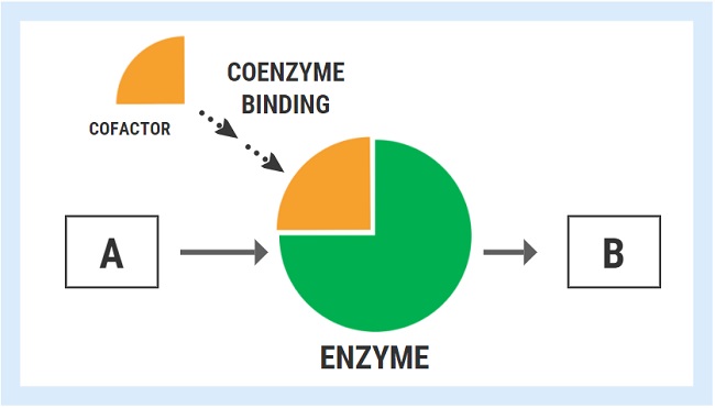
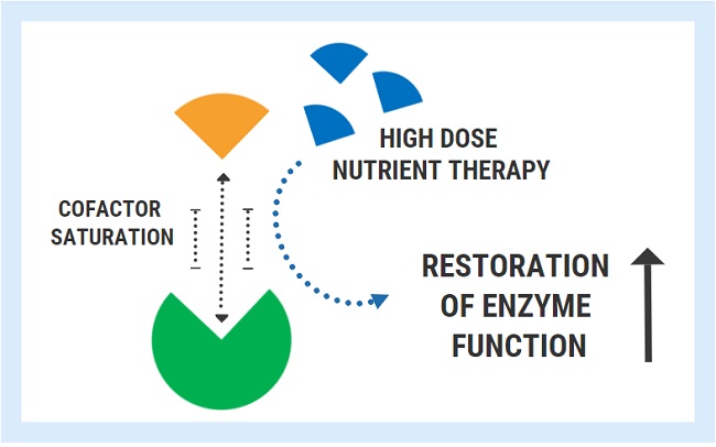
I learned that genes drive enzymes that do all the myriad tasks to run our bodies (which don’t just function automatically), and that certain vitamins and minerals are required to assist the enzymes, as specific “cofactors.” Genetic SNPs require even more nutritional support than is normal to help enzymes function better. So my focus shifted toward using basic vitamins and minerals to support my genetic impairments. I now understood that I needed extra B12, folate, glutathione, and more. I began following Ben Lynch’s work in elucidating the MTHFR genetic issue, as I had MTHFR A1298C.

Also in 2013, given my struggle with diarrhea, I was diagnosed with microscopic colitis via a biopsy with colonoscopy. In 2014, I learned about small intestinal bacterial overgrowth (SIBO), which gave me a more specific understanding of my “gut infections,” and tested positive for methane SIBO. I worked with a SIBO-oriented practitioner on specific herbal treatments with some short-lived success.

At the end of 2014, I learned that I have Ehlers Danlos Syndrome (EDS, Hypermobile Type), confirmed by a specialist. I came to understand that my “bendiness” likely had implications in terms of chronic illness, and I saw my bunion and carpal tunnel surgeries in a new context, as part of this syndrome.

Even with these breakthroughs in understanding, I still relentlessly searched deeper for root causes.

Genetic Kinetics

In 2018, Ben Lynch published Dirty Genes, focusing on a number of common yet impactful SNPs.

I learned that I had NEARLY ALL of these SNPs – NEARLY ALL as “doubles” and even a “deletion.” (Deletions are worse than doubles; doubles are worse than singles.) Researching further, I had doubles in many related genes with added interactive impacts. Typically people might have just a few of these SNPs.

Understanding my “dirty gene” SNPs revealed that I could be deficient in methylation, detoxification, choline synthesis, nitric oxide synthesis, neurotransmitter processing, and histamine processing. Each of these SNPs could potentially impact sleep, digestion, and much more in numerous ways. Now I potentially had a myriad of root causes.

Lynch warns people to clean up their health act before supplementing the cofactors, whereas I’d cleaned mine up years prior. Sadly, I found only limited improvements in adding his nutritional protocol. Suffice it to say I felt rather overwhelmed and disheartened.

But at the same time, I gained vital and necessary insights. I now understood why I had mercury poisoning twice: detox impairments. I understood why I had Raynaud’s, chilblains, and poor circulation: nitric oxide impairments. My migraines could be histamine overload. I needed high levels of choline for the PEMT gene to prevent fatty liver disease and SAMe for the COMT gene. Much was yet still unexplained. So I relentlessly soldiered on, following every lead, clue, and a new piece of information.

Later in 2018, a friend who also has EDS encouraged me to learn about Mast Cell Activation Syndrome (MCAS), as many with EDS also have this condition. A few weeks later, I had a three-day flare of many issues, which prompted me to delve into the MCAS world, which was just as complex as the genetic approach. In working with an MCAS specialist, I honed in on three supplements, quercetin, palmitoylethanolamide, and luteolin, to help stabilize mast cells, which improved my bladder pain, bone pain, migraines, fatigue, and generalized pain. This was the culmination of months of research and work. All of this points to further genetic involvement, even though I lack specifics.

Downward Spiral

Twenty-nineteen brought further insights. I integrated circadian rhythm entrainment work. I tried a low-sulfur diet, suspecting hydrogen sulfide SIBO, which made me feel worse; and I began taking dietary oxalates somewhat more seriously after testing positive on a Great Plains OAT test. I did glyphosate and toxicity testing, which provided a picture of my toxic load. Testing also indicated high oxidative stress and mitochondrial issues (very interrelated). Hair Tissue Mineral Analysis (HTMA) testing, with the assistance of a specialist, helped me understand my mineral status and to begin rebalancing and repleting.

In 2020, I took a hiatus from all this effort, during which time I turned my attention towards personal matters, but 2021 has been a doozy in redoubling my health efforts. My digestion had worsened, so I focused on this area. I learned about sucrase-isomaltase deficiency, a lack of certain enzymes to digest sucrose and starch. I hadn’t tolerated sugar and starch for years, and I found I had a SNP for this condition. In January, a zero-carb trial diet helped me feel much better, so I continued. I tested positive for hydrogen sulfide SIBO, and I wrestled with this “whole-other-SIBO-beast” – in February trying again the low-sulfur diet and again feeling worse. Combining the zero-starch and low-sulfur diets left few options. Despite all my best efforts, I experienced a downward spiral with a loss of appetite, nausea, and vomiting every few days.

Discovering Thiamine

Around this time, I read an article about low thiamine (Vitamin B1) lowering intracellular potassium – I had been trying unsuccessfully to raise my potassium level in my HTMA work. I began following author Elliot Overton’s articles and videos on thiamine deficiency and oxalates. I was finally persuaded to take oxalates seriously. I then read the definitive book “Thiamine Deficiency Disease, Dysautonomia, and High Calorie Malnutrition” by Drs. Derrick Lonsdale and Chandler Marrs. I learned how B1 was key in many processes involving energy, digestion, and much more. I found that I had multiple SNPs in the B1-dependent transketolase gene, which is pivotal in several pathways. I gained some understanding of how all this related to some of my other genetic impairments, and why I might need high dose thiamine to overcome some issues.

All this was quite a revelation for me. It fit perfectly with my emphasis on vitamins and minerals to assist genes…but why hadn’t I learned of B1’s significance sooner?

In early March, I began my thiamine odyssey with 100 mg of thiamine HCL, upping the dose every couple of days. At 300mg HCL, I added 50 mg of TTFD, a more potent and bioavailable form of B1, then continued to up the TTFD dose every few days.

Similar to my experience with other vitamins, I was able to proceed rather quickly in dose increases. Many other people are not so fortunate and must go much more slowly. I already had in place most of thiamine’s cofactors (such as glutathione, other B vitamins, and methylation support) – so perhaps this helped me proceed more readily. Without these cofactors, peoples’ thiamine efforts often fail.

Magnesium is one of the most important thiamine cofactors, and for me, the most challenging. My gut cannot handle it, so I must apply it transdermally two or more times a day. At times, I had what I interpreted as low magnesium symptoms: racing and skipping heart, but these resolved as I continued.

Additionally, one must be prepared for “paradoxical reactions.” Worse-before-better symptoms hit me the day after thiamine dose increases: gut pain, sour stomach, headache, fatigue, and soreness.

My symptoms improved as I increased the dosing. When I added 180 mg of benfotiamine early on, my bit of peripheral neuropathy immediately cleared. This form of B1 helps nerve issues. As I increased my thiamine dosing, the nausea abated, my appetite came roaring back, and gastritis disappeared. Diarrhea, fatigue, and restless legs improved. I was able to jog again. My digestion improved without trying to address the SIBO and inflammation directly; the strict keto and low oxalate diets may have also helped.

In June, I attained a whopping TTFD dose of 1500 mg but did not experience further resolution beyond 1200mg, so I dropped back down. At 1200mg for a month, a Genova NutrEval test revealed that I was not keeping pace with TTFD’s needed cofactors, especially glutathione and its substrates. Not too surprising, given my malabsorption issues and my already high need for these nutrients. I dropped the TTFD to 300 mg, but quickly experienced fatigue. I’m now at 750 mg, which is still a large dose, and clearly, there is more to my situation than thiamine can address. I still have diarrhea and insomnia, and continue working to address these.

The Next Chapter

With TTFD, its cofactors, and my new gains in place, I’ve turned my attention towards a duo of genetic deletions that I have in GPX1 (glutathione peroxidase 1, one of Lynch’s dirty genes) and CAT (catalase). Both of these enzymes break down hydrogen peroxide (H2O2), a byproduct of numerous bodily processes. This unfortunate double-whammy causes me a build-up of damaging H2O2 and lipid peroxides – in other words, oxidative stress, a major factor in mitochondrial impairment, many diseases, and aging. This might be one of my biggest and yet-unaddressed issues, and I am digging deep into the published medical literature. This new chapter is currently unfolding.

I believe these two deletions are related to my father’s radiation exposure, for reasons beyond the scope of this article. But what about all the other SNPs? Many questions remain unanswered.

All my gains have been so hard-won, involving much research, effort, and supplementation. Yet what other options do I have, besides playing whack-a-mole and spiraling downward? Looking back, my improvements have been substantial, given the multitude of issues I’ve had to deal with. Perhaps now at 60, my life can start to open again to more than just self-care.

I hate to think of where I would be now, had I never come across the thiamine deficiency issue. I believe a number of factors had driven my thiamine status dangerously low earlier this year, such as malabsorption, oxidative stress, and hydrogen sulfide SIBO. I’m forever grateful to Lonsdale, Marrs, and Overton for their invaluable thiamine work that helped guide me back from the brink, and to the numerous doctors and practitioners who have helped me get this far. Perhaps my story can help others struggling with chronic health issues.

We Need Your Help

More people than ever are reading Hormones Matter, a testament to the need for independent voices in health and medicine. We are not funded and accept limited advertising. Unlike many health sites, we don’t force you to purchase a subscription. We believe health information should be open to all. If you read Hormones Matter, like it, please help support it. Contribute now.

Yes, I would like to support Hormones Matter.

Image credit: First U.S. nuclear field exercise conducted on land; troops shown are a mere 6 miles from the blast. Nevada Test Site, 1 November 1951. This image is a work of a U.S. military or Department of Defense employee, taken or made as part of that person’s official duties.

As a work of the U.S. federal government, the image is in the public domain in the United States.

This article was published originally on September 23, 2021.

SIBO, IBS, and Constipation: Unrecognized Thiamine Deficiency?

158447 views

In many of my clients, chronic upper constipation and gastroesophageal reflux disease (GERD) are misdiagnosed as bacterial overgrowth. Unfortunately, they are often non-responsive to antimicrobial treatments. Yet, sometimes the issues are fixed within a few days of vitamin B1 repletion. This has shown me that often times, the small intestinal bacterial overgrowth (SIBO) is simply a symptom of an underlying vitamin B1 or thiamine deficiency.

GI Motility and Thiamine

The gastrointestinal (GI) tract is one of the main systems affected by a deficiency of thiamine. Clinically, a severe deficiency in this nutrient can produce a condition called “Gastrointestinal Beriberi”, which in my experience is massively underdiagnosed and often mistaken for SIBO or irritable bowel syndrome with constipation (IBS-C). The symptoms may include GERD, gastroparesis, slow or paralysed GI motility, inability to digest foods, extreme abdominal pain, bloating and gas. People with this condition often experience negligible benefits from gut-focused protocols, probiotics or antimicrobial treatments. They also have a reliance on betaine HCL, digestive enzymes, and prokinetics or laxatives.

To understand how thiamine impacts gut function we have to understand the GI tract. The GI tract possesses its own individual enteric nervous system (ENS), often referred to as the second brain. Although the ENS can perform its job somewhat autonomously, inputs from both the sympathetic and parasympathetic branches of the autonomic nervous system serve to modulate gastrointestinal functions. The upper digestive organs are mainly innervated by the vagus nerve, which exerts a stimulatory effect on digestive secretions, motility, and other functions. Vagal innervation is necessary for dampening inflammatory responses in the gut and maintaining gut barrier integrity.

The lower regions of the brain responsible for coordinating the autonomic nervous system are particularly vulnerable to a deficiency of thiamine. Consequently, the metabolic derangement in these brain regions caused by deficiency produces dysfunctional autonomic outputs and misfiring, which goes on to exert detrimental effects on every bodily system – including the gastrointestinal organs.

However, the severe gut dysfunction in this context is not only caused by faulty central mechanisms in the brain, but also by tissue specific changes which occur when cells lack thiamine. The primary neurotransmitter utilized by the vagus nerve is acetylcholine. Enteric neurons also use acetylcholine to initiate peristaltic contractions necessary for proper gut motility. Thiamine is necessary for the synthesis of acetylcholine and low levels produce an acetylcholine deficit, which leads to reduced vagal tone and impaired motility in the stomach and small intestine.

In the stomach, thiamine deficiency inhibits the release of hydrochloric acid from gastric cells and leads to hypochlorydria (low stomach acid). The rate of gastric motility and emptying also grinds down to a halt, producing delayed emptying, upper GI bloating, GERD/reflux and nausea. This also reduces one’s ability to digest proteins. Due to its low pH, gastric acid is also a potent antimicrobial agent against acid-sensitive microorganisms. Hypochlorydria is considered a key risk factor for the development of bacterial overgrowth.

The pancreas is one of the richest stores of thiamine in the human body, and the metabolic derangement induced by thiamine deficiency causes a major decrease in digestive enzyme secretion. This is one of the reasons why those affected often see undigested food in stools. Another reason likely due to a lack of brush border enzymes located on the intestinal wall, which are responsible for further breaking down food pre-absorption. These enzymes include sucrase, lactase, maltase, leucine aminopeptidase and alkaline phosphatase. Thiamine deficiency was shown to reduce the activity of each of these enzymes by 42-66%.

Understand that intestinal alkaline phosphatase enzymes are responsible for cleaving phosphate from the active forms of vitamins found in foods, which is a necessary step in absorption. Without these enzymes, certain forms of vitamins including B6 (PLP), B2 (R5P), and B1 (TPP) CANNOT be absorbed and will remain in the gut. Another component of the intestinal brush border are microvilli proteins, also necessary for nutrient absorption, were reduced by 20% in the same study. Gallbladder dyskinesia, a motility disorder of the gallbladder which reduces the rate of bile flow, has also been found in thiamine deficiency.

Malnutrition Induced Malnutrition

Together, these factors no doubt contribute to the phenomena of “malnutrition induced malnutrition”, a term coined by researchers to describe how thiamine deficiency can lead to all other nutrient deficiencies across the board. In other words, a chronic thiamine deficiency can indirectly produce an inability to digest and absorb foods, and therefore produce a deficiency in most of the other vitamins and minerals. In fact, this is indeed something I see frequently. And sadly, as thiamine is notoriously difficult to identify through ordinary testing methods, it is mostly missed by doctors and nutritionists. To summarize, B1 is necessary in the gut for:

  • Stomach acid secretion and gastric emptying
  • Pancreatic digestive enzyme secretion
  • Intestinal brush border enzymes
  • Intestinal contractions and motility
  • Vagal nerve function

Based on the above, is it any wonder why thiamine repletion can radically transform digestion? I have seen many cases where thiamine restores gut motility. Individuals who have been diagnosed with SIBO and/or IBS and are unable to pass a bowel movement for weeks at a time, begin having regular bowel movements and no longer require digestive aids after addressing their thiamine deficiency. In fact, the ability of thiamine to address these issues has been known for a long time in Japan.

TTFD and Gut Motility

While there are many formulations of thiamine for supplementation, the form of thiamine shown to be superior in several studies is called thiamine tetrahydrofurfuryl disulfide or TTFD for short. One study investigated the effect of TTFD on the jejunal loop of non-anesthetized and anesthetized dogs. They showed that intravenous administration induced a slight increase in tone and a “remarkable increase” in the amplitude of rhythmic contractions for twenty minutes. Furthermore, TTFD applied topically inside lumen of the intestine also elicited excitation.

Another study performed on isolated guinea pig intestines provided similar results, where the authors concluded that the action of TTFD was specifically through acting on the enteric neurons rather than smooth muscle cells. Along with TTFD, other derivatives have also been shown to influence gut motility. One study in rats showed an increase in intestinal contractions for all forms of thiamine including thiamine hydrochloride (thiamine HCL), S-Benzoyl thiamine disulphide (BTDS -a formulation that is  somewhat similar to benfotiamine), TTFD, and thiamine diphosphate (TPD). A separate study in white rats also found most thiamine derivatives to be effective within minutes.

Most interestingly, in another study, this time using mice, the effects of thiamine derivatives on artificially induced constipation by atropine and papaverine was analyzed. The researchers tested whether several thiamine derivatives could counteract the constipation including thiamine pyrophosphate (TPP), in addition to the HCL, TTFD and BTDS forms. Of all the forms of thiamine tested, TTFD was the ONLY one which could increase gut motility. Furthermore, they ALSO showed that TTFD did not increase motility in the non-treatment group (non-poisoned with atropine). This indicated that TTFD did not increase motility indiscriminately, but only when motility was dysfunctional. Finally, severe constipation and gastroparesis identified in patients with post-gastrectomy thiamine deficiency, was alleviated within a few weeks after a treatment that included three days of IV TTFD at 100mg followed by a daily dose of 75mg oral TTFD. Other symptoms also improved, including lower limb polyneuropathy.

To learn more about how thiamine affects gut health:

We Need Your Help

More people than ever are reading Hormones Matter, a testament to the need for independent voices in health and medicine. We are not funded and accept limited advertising. Unlike many health sites, we don’t force you to purchase a subscription. We believe health information should be open to all. If you read Hormones Matter, like it, please help support it. Contribute now.

Yes, I would like to support Hormones Matter. 

Photo by Johannes Krupinski on Unsplash.

This article was first published on HM on June 1, 2020. 

How Can Something As Simple as Thiamine Cause So Many Problems?

162563 views

I have read a criticism that thiamine deficiency is “too simple” to explain the devastating nature of the post Gardasil illnesses or the systemic adverse reactions to some medications. Sometimes, it is the simple and overlooked elements that are the most problematic.

Understanding Thiamine’s Role in Complex Adverse Reactions – The Limbic System

The lower part of the brain, called the brainstem, is a like computer, controlling the most basic aspects of survival, from breathing and heart rate, hunger and satiety, to fight or flight and reproduction. This computer-like function within the brainstem is called the autonomic system (ANS). The ANS together with the limbic system act in concert to regulate our most basic survival functions and behaviors. Both require thiamine to function.

Postural Orthostatic Tachycardia Syndrome or POTS , a type of dysautonomia (dysregulation of the autonomic system) seems to be the among the commonest manifestations of the Gardasil effect. Many cases have been diagnosed already, while others present all of the symptoms but have yet to receive a diagnosis. Dysautonomia and POTS have also been observed with adverse reactions to other medications, as well. Dysautonomia and POTS, at the most basic level, represent a chaotic state of the limbic-autonomic system. Let me explain.

Fragmented Fight or Flight

The brainstem autonomic system together with the limbic system enable us to adapt to our environment, presiding over a number of reflexes that allow us to survive. For example, fight-or-flight is a survival reflex, triggered by perception of a dangerous incident that helps us to kill the enemy or escape. This kind of “stress event” in our ancestors was different from that we experience today. Wild animal predators have been replaced by taxes/business deadlines/rush hour traffic etc. These are the sources of modern stress. The beneficial effect is that the entire brain/body is geared to physical and mental response. However, it is designed for short term action and consumes energy rapidly. Prolonged action is literally exhausting and results in the sensation of fatigue. In the world of today where dietary mayhem is widespread, this is commonly represented as Panic Attacks, usually treated as psychological. They are really fragmented fight-or-flight reflexes that are triggered too easily because of abnormal brain chemistry.

Thiamine and Oxidative Metabolism: The Missing Spark Plug

Our brain computers rely completely on oxidative metabolism represented simply thus:

Fuel + Oxygen + Catalyst = Energy

Each of our one hundred trillion body/brain cells is kept alive and functioning because of this reaction. It all takes place in micro “fireplaces” known as mitochondria. Oxygen combines with fuel (food) to cause burning or the combustion – think fuel combustion engine. We need fuel, or gasoline, to burn and spark plugs to ignite in order for the engines to run.

In our body/brain cells it is called oxidation. The catalysts are the naturally occurring chemicals we call vitamins (vital to life). Like a spark plug, they “ignite” the food (fuel). Absence of ANY of the three components spells death.

Antioxidants like vitamin C protect us from the predictable “sparks” (as a normal effect of combustion) known as “oxidative stress”.  Vitamin B1, is the spark plug, the catalyst for these reactions. As vitamin B1, thiamine, or any other vitamin deficiency continues, more and more damage occurs in the limbic system because that is where oxygen consumption has the heaviest demand in the entire body. This part of the brain is extremely sensitive to thiamine deficiency.

Why Might Gardasil Lead to Thiamine Deficiency?

We do not know for sure how Gardasil or other vaccines or medications have elicited thiamine deficiency, but they have. We have two girls and one boy, tested and confirmed so far. More testing is underway. Thiamine deficiency in these cases may not be pure dietary deficiency. It is more likely to be damage to the utilization of thiamine from as yet an unknown mechanism, affecting the balance of the autonomic (automatic) nervous system. It is certainly able to explain POTS (one of the many conditions that produce abnormal ANS function) in two Gardasil affected girls. Beriberi, the classic B1 deficiency disease, is the prototype for ANS disease. Administration of thiamine will not necessarily bring about a cure, depending on time since onset of symptoms, but it may help.

Thiamine Deficiency Appetite and Eating Disorders

Using beriberi as a model, let us take appetite as an example of one of its many symptoms. When we put food into the stomach, it automatically sends a signal to a “satiety center” in the computer. As we fill the stomach, the signals crescendo and the satiety center ultimately tells us that we have eaten enough. Thiamine deficiency affects the satiety center, wrecking its normal action. Paradoxically it can cause anorexia (loss of appetite) or the very opposite, a voracious appetite that is never satisfied and may even go on to vomiting. It can also shift from anorexia to being voracious at different times within a given patient. That is why Anorexia Nervosa and Bulimia represent one disease, not two.

Thiamine Deficiency, Heart Rate and Breathing

The autonomic nervous system, responsible for fight or flight, regulates heart activity, accelerating or decelerating according to need. So heart palpitations are common in thiamine deficiency. Its most vital action is in control of automatic breathing and thiamine deficiency has long been known to cause infancy sudden death from failure of this center in brainstem.

Thiamine Deficiency and Sympathetic – Parasympathetic Regulation

The hypothalamus is in the center of the brain computer and it presides over the ANS, as well as the endocrine (hormone) system. The ANS has two channels of communication known as sympathetic (governs action) and parasympathetic (governs the body mechanisms that can be performed when we are in a safe environment: e.g. bowel activity, sleep, etc.). When the ANS system is damaged, sometimes by genetic influence, but more commonly by poor diet (fuel), our adaptive ability is impaired. A marginal energy situation might become full blown by a stress factor. In this light, we can view vaccines and medications as stress factors. From false signal interpretation, we may feel cold in a warm environment, exhibiting “goose bumps on the skin”, or we may feel hot in a cold environment and experience profuse sweating. The overriding fatigue is an exhibition of cellular energy failure in brain perception.

Sometimes, it really is the simple, overlooked, elements that cause the most devastating consequences to human health. Thiamine deficiency is one of those elements.

To learn more about thiamine testing: Thiamine Deficiency Testing: Understanding the Labs.

We Need Your Help

More people than ever are reading Hormones Matter, a testament to the need for independent voices in health and medicine. We are not funded and accept limited advertising. Unlike many health sites, we don’t force you to purchase a subscription. We believe health information should be open to all. If you read Hormones Matter, like it, please help support it. Contribute now.

Yes, I would like to support Hormones Matter. 

Photo by Robina Weermeijer on Unsplash.

This article was published originally in October 2013.

Rest in peace Derrick Lonsdale, May 2024.


Thiamine, Epigenetics, and the Tale of the Traveling Enzymes

10740 views

For some time now, we have been covering all the ways in which thiamine deficiency influences disease. The primary mechanisms are through the down regulation of mitochondrial enzymes critical for ATP production. The lack of thiamine impairs mitochondrial functioning significantly leading to complex, debilitating, chronic, and sometimes, deadly illnesses. With mitochondrial energy a requisite for cell functioning and survival it is easy to see how the diminishment of mitochondrial functioning would negatively impact health and how high-energy physiological systems like the nervous and cardiovascular systems might be particularly hard hit. More than just just derailing cell function, as if that wasn’t problematic enough, when mitochondrial energy production slows, the adaptive cascades that ensue include epigenetic modification, not only at the level of mitochondrial DNA (mtDNA), but also, at the level of gene expression from the cell nucleus or nDNA. This is a huge discovery with broad implications about health and disease. It means that all sorts of things considered innocuous, are directly influencing gene activation and deactivation by way of the mitochondria.

Epigenetics: How the Cells Adapt to the Environment

Epigenetic modification refers to the activation or deactivation of chromosomal gene expression absent mutation. These changes can be heritable and often are. Strictly speaking, epigenetics involves changes in methylation, histone modification and/or alterations in non-coding RNA that affect transcription. Epigenetics is the way our genome adapts to environmental circumstances and prepares our offspring to do the same. The majority of epigenetic work focuses on genomic changes. That is, those variables that affect gene expression from the cell nucleus or nDNA. There is a growing body of evidence, however, that mitochondrial DNA (mtDNA) are affected by epigenetic factors in much the same way as nDNA. In fact, given the mitochondria’s role in cell survival, one might suspect that mitochondria are more susceptible to environmental epigenetics, perhaps even the first responders and/or the initiators of chromosomal genetic changes.

Considering that nDNA accounts for over 90% of the proteins involved in mitochondrial functioning, how could damaged mitochondria, even functionally inefficient mitochondria, not affect gene expression in the cell’s nucleus? Though we tend to think of mitochondria as self-contained and discrete entities, black boxes of sorts, where stuff goes in and ATP magically comes out, this is not only not the case, it seems biologically illogical to think that way. So much of mitochondrial functioning is related to its environmental milieu. In fact, increasingly researchers are finding that the mitochondria are the center of the organismal universe, sensing and signaling danger, and effectively, regulating all adaptive responses, including epigenetic modification of the proteins encoded by the cell nucleus.

Beyond the strict definition of epigenetics, it seems to me that any factor that altered mitochondrial function, would eventually alter gene expression by any number of mechanisms, not just the direct ones. That is, because the mitochondria produce the energy required for cell survival, anything that derails their capacity to produce ATP, pharmaceutical and environmental chemicals, for example, should be considered, ipso facto, epigenetic modulators. Energy is a fundamental requirement for life. How could energy depletion not affect gene expression? It does, and now we know how.

Starve the Mitochondria, Alter the Genome

It turns out, when the mitochondria are starving and/or damaged the key enzyme complex that sits atop the entire energy production pathway and is critical for the production of the substrates involved directly (yes, I said directly) with gene expression, migrates from the mitochondria across the cell and into the cell’s nucleus where it then sets up shop and begins its work there. Sit with that for a minute. The entire enzyme complex decides that things are not working where it is, so it hitches a ride with some transporter proteins, several of them along the way, to find a more suitable home. The enzyme complex ‘knows’ that its functions are critical for survival and so it must move or die. What an incredible bit of adaptive capacity. A symbiosis, if you will.

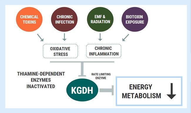
To make matters even more interesting, the traveling enzyme complex just so happens to be the pyruvate dehydrogenase complex (PDC) and you guessed it, the PDC is highly, and I mean highly, thiamine dependent. But wait, there’s more. Several other enzymes involved in mitochondrial bioenergetics are also thiamine dependent. These include: alpha-ketoglutarate dehydrogenase (α- KGDH) in the tricarboxylic acid (TCA) or citric acid cycle, transketolase (TKT) within the pentose phosphate pathway (PPP), the branched chain alpha-keto acid dehydrogenase complex (BCKDC) involved in amino acid catabolism and, more recently, thiamine has be identified as a co-factor in fatty acid metabolism via an enzyme called 2-hydroxyacyl-CoA lyase (HACL1) in the peroxisomes (organelles that break down fatty acids before transporting them to the mitochondria). So thiamine deficiency is problematic. It not only causes dysfunction in the mitochondria, reducing bioenergetic capacity, but if severe enough and/or chronic, it alters the genome. And those changes are likely heritable. That is, your thiamine deficiency likely will affect your children’s ability to process thiamine.

Thiamine Deficiency and Gene Expression

Without sufficient thiamine, the PDC enzyme complex does not function well and because of its geographic position at the entry point into the citric acid cycle, when it is not working at capacity, everything below it eventually grinds to a halt resulting in severe neurological and neuromuscular deficits. Absent congenital PDC disorders, however, when it simply is inefficient or starved for its cofactors, metabolic disorders ensue because we cannot convert carbohydrates into ATP and the sugars that normally would be converted into energy, remain unmetabolized, floating around outside the cells and causing the whole cascade of effects that mark type 2 diabetes. When the PDC is inefficient, ATP levels wane and fatigue ensues. Systems that are highly energy dependent are hit the hardest. Think brain, heart, muscles, GI tract. When mitochondria are inefficient or damaged, reactive oxygen species (ROS) increase. Anti-oxidant capacity decreases and further damage to the PDC ensues.  Inflammation increases, immune function decreases. Cell level hypoxia grows. Alternative energy pathways are activated, those that are endemic of cancer. Yes, cancer can be considered a metabolic disorder.

When the PDC is inefficient, mtDNA heteroplasmy, the balance between mutated mtDNA and healthy mtDNA grows with each mitogenic cycle. This, of course, further derails mitochondrial capacity (and increases the need for thiamine). Absent available resources, mitochondrial death cascades are initiated. And now, we know at some point in this disease and death spiral, when ATP diminishes and the litany of adaptive measures fail to maintain sufficient energy availability, the PDC up and leaves its mitochondrion and sets up shop in the cell nucleus, in what is presumably a last ditch effort to save its cell and the organism as a whole. What a remarkable bit of adaptive capacity.

The researchers, who discovered this, ruled out the possibility that the PDC enzyme complex was already in the cell nucleus. It wasn’t. It traveled by way of several transporter proteins. They also found that once in the nucleus, it began producing acetyl-coenzyme A (CoA). Acetyl-CoA is requisite for the acetylation and deacetylation of histones, post translational changes in DNA, but also lysine acetylation, which further affects metabolism – mitochondrial energy homeostasis. With insufficient acetyl-CoA and insufficient acetylation, DNA replication is aberrant. Moreover, without sufficient acetyl-CoA, acetylcholine synthesis, an incredibly important transmitter for nervous system functioning, diminishes.

A Constitutively Active Enzyme: What Could Possibly Go Wrong?

Even more interesting, the PDC in the cell nucleus appears to be constitutively active. Unlike in healthy mitochondria where checks and balances prevail, when the PDC travels to the nucleus it has no feedback control to temper its activity. The enzymes that normally shut down PDC production (pyruvate dehyrodgenase kinase) in the mitochondria, were not present in the cell nucleus, and thus, once the PDC was turned on, it stayed on. That means when the PDC translocates to the cell nucleus it operates independently much like a Warburg effect in cancer metabolism; one that fully supplants healthier metabolic pathways. Can you say tumorogenesis?

This research has enormous implications for everything from cancer to Alzheimer’s disease and everything in between that involves metabolic disturbances like diabetes and heart disease. Metabolism begins and ends with the mitochondria, at the PDC, and the PDC is highly dependent on thiamine. Every pharmaceutical and environmental chemical damages the mitochondria in some manner or another and that damage inevitably reduces ATP capacity. Some chemicals also deplete thiamine directly, thus downregulating the activity of the PDC and the other thiamine dependent enzymes. Even when these chemicals don’t deplete thiamine directly, the western diet is more often than not thiamine deficient, sometimes only marginally, others time quite significantly so with symptoms already expressed, but very rarely recognized. We have known that thiamine was critical for health and survival for some time. Now we have one more reason to tread carefully with lifestyles and exposures that deplete thiamine.

We Need Your Help

More people than ever are reading Hormones Matter, a testament to the need for independent voices in health and medicine. We are not funded and accept limited advertising. Unlike many health sites, we don’t force you to purchase a subscription. We believe health information should be open to all. If you read Hormones Matter, like it, please help support it. Contribute now.

Yes, I would like to support Hormones Matter. 

David Goodsell, CC BY 3.0, via Wikimedia Commons

This article was first published on April 25, 2017. 

Mystery Illness: You Are Not Alone

13191 views

Hormones Matter is a health oriented website edited by Chandler Marrs, PhD. She has long recognized the need for people to report their “mystery illnesses”, simply because they are slipping through the cracks in modern medicine. My association with Dr. Marrs is a very fruitful one because we both have the same viewpoint. This viewpoint embraces the concept that the present disease model is antiquated and badly needs to be revised. In a recent post, I have defined what we mean by a “medical model”. We both have found that a common health problem, largely unrecognized for its true cause, is a polysymptomatic illness that is almost invariably labeled psychosomatic. I will try to explain.

Food, Energy, and Illness

Much of our food is broken down to glucose, the primary fuel of the brain. This has given rise to a common concept that taking virtually any form of sugar is a way to develop “quick energy”. Before the processing of sugar in the body was understood, athletes would sometimes load up on it. We now know that this defeats the purpose. Very much like a car where an excess of gasoline “chokes” the engine, an excess of sugar has a very similar effect, particularly in the brain. An additional effect of sugar is the extremely sweet taste that sends a signal from the tongue to centers in the brain that gives the person an extreme sense of pleasure. It has been shown in animal studies that sugar is more addictive than cocaine and a book was published in 1973 entitled “Sweet and Dangerous”. The author, Dr. John Yudkin, was a professor of nutritional studies in a major London hospital. He was able to show that sugar was the cause of many modern diseases. It is indeed hard for people to understand that such an appreciated delight is dangerous to our health. If we turn to nature, you will find that sugar is never found in its free state. It is always found in fruit and vegetables where fiber is a vital component in its processing. The sweet taste from eating a banana or an orange is the way that Mother Nature designed it and it is a healthy way of experiencing a sweet taste.

Glucose is burned (oxidized) in cellular “engines” (mitochondria) and it is a very complex process. The net result is energy that is stored in a chemical substance known as adenosine triphosphate (ATP). The nearest analogy would be a battery because the energy that drives all our mental and physical functions is electrical in nature.

By far and away the commonest personal story posted on Hormones Matter is a polysymptomatic illness that is the result of inefficient energy transduction and its major effect is in the brain. To put it as simply as possible, food is not being converted into energy in sufficient amount to meet the stresses of merely being alive. The most susceptible part of the brain that is affected is the part that controls our ability to adapt to living in an environment that is essentially hostile. Using a specialized nervous system and a bunch of glands that produce hormones, this part of the brain signals every organ in the body to participate. Now obviously, if no energy were produced we would die and that is indeed a major cause of death. However this common polysymptomatic illness affecting so many people is based on an inefficient energy production, not a complete lack. It can vary in its degree of severity depending on nutritional and genetic factors. The dominant effect is “psychological”, symptoms such as undue fatigue, depression, anxiety and anger. It can run the gamut of our emotional reactions. In fact, because of its emotional implications, I have suggested that the common state of violence in America is a reflection of our uncontrolled hedonism. Can a person nursing a perceived grievance become violent if the emotional controls are too easily activated?

Energy lack is quickly recognized as dangerous by the brain. It causes a sense of panic to be felt by the affected person. That is why “panic attacks” have been recognized incorrectly as a “psychological disease that requires a medicine to tranquilize the patient” whereas they really represent a fight-or-flight reflex, naturally designed to get the affected person “out of perceived danger, i.e. energy deficiency”. The affected person seeks medical help, but this effect in the brain is seen by most physicians as “psychological”, as though the patient is inventing the symptoms. The diagnosis is, “it’s all in your head”. The irony is that although the symptoms are indeed the result of a function “in the head”, they are evidence of a sick brain lacking in adequate energy and therefore have an understandable origin and meaning. Also, the symptoms are easily erased by administration of non-caloric nutrient supplements when they are initially experienced. If allowed to continue unchecked, sometimes for years, they may lead to the irreversible damage characterized as a neurodegenerative disease.

Because the dominant effect is in the part of the brain that controls the specialized nervous system, it begins to send out exaggerated “panic” signals to the organs of the body. The result is a variable assortment of physical effects— heart palpitations, breathing problems, diarrhea, often alternating with constipation, whole body pain, migraine headaches, nasal congestion, nausea with or without vomiting, chest or abdominal pain, pins and needles etc. In other words, any organ in the body may be activated or non-activated because the pattern of our adaptive mind/body machinery is adversely affected. The very important point is this: each and every action of the brain/body union requires energy, even sleep!

Perhaps the most common symptom is severe fatigue and this has given rise to a common diagnosis of Chronic Fatigue Syndrome (CFS). It is worth noting that it is often associated with Irritable Bowel Syndrome (IBS) and it seems to be medically accepted that two diseases, both of “unknown cause” can occur in a patient at the same time. That seems to be a product of illogical thinking based on the present medical model.

Share Your Story

Anyone encountering this website is encouraged to write his or her health story and share it as a blog post. These stories help raise awareness about the scope of illnesses affecting us all and add to the knowledge base. To share your health story, send us a note here.

If you have specific questions about health and illness, we recommend that you “surf” the site because there are many posts on a variety of topics with long and detailed comment threads, one or more of which may be similar to your own story and may answer your questions.

We Need Your Help

More people than ever are reading Hormones Matter, a testament to the need for independent voices in health and medicine. We are not funded and accept limited advertising. Unlike many health sites, we don’t force you to purchase a subscription. We believe health information should be open to all. If you read Hormones Matter, like it, please help support it. Contribute now.

Yes, I would like to support Hormones Matter. 

Image by Leandro De Carvalho from Pixabay .

This article was published originally on December 2, 2019. 

Rest in peace Derrick Lonsdale, May 2024.

Oxalate Degrading Microbes: Reconsidering Pathogenesis

41033 views

An exciting new paper was published recently which should turn the oxalate world upside down. The authors hunted public databases and compiled a list of microbes that possess variations of a gene called oxalyl CoA decarboxylase. That gene makes a protein that degrades oxalate.

Oxalate is an ion used by microbes for communication between fungus and bacteria, but it is found at varying levels in plant food. Our bodies also manufacture oxalate during a stress response. Everybody makes some amount of oxalate in their bodies and eats sometimes huge amounts of oxalate in foods like spinach, beets, nuts and seeds, and also (alas!) chocolate. When oxalate in our bodies gets too high, it causes dysbiosis and becomes dangerous, tangling with mineral metabolism, and delivering harsh changes to mitochondrial function, adding in distressing levels of oxidative stress.

Humans lack any oxalate-degrading gene in our own genome. That means we cannot degrade oxalate using our own talents. Instead, we are reliant upon our microbes in the gut to degrade oxalate. From studying isolated human beings who have never seen an antibiotic, scientists have realized that like them, our ancestors had plenty of oxalate degrading microbes in the gut (termed oxalotrophic microbes). The job of these microbes was to keep oxalate from food from getting into the inside of their bodies. That system worked well even when the diet was high in oxalate, but things changed when our microbiome began to be damaged by antibiotics and antifungals, and other hits.

In this discussion, I would like to examine findings from this recently published study from China:  Abundance, Functional, and Evolutionary Analysis of Oxalyl-Coenzyme A Decarboxylase in Human Microbiota where they identified the microbial species that possess one of the two genes that we know are used to degrade oxalate in our intestines. We will also examine some other issues that this new knowledge unwraps.

Oxalate Degradation Is Dependent upon Thiamine and Is Impaired by Antibiotics

The protein oxalyl coenzyme A decarboxylase is dependent upon thiamine. It requires thiamine that is in other forms to be converted into thiamine diphosphate (TPP). Scientists have told us that this is the primary form of thiamine that microbes will make and use.

Some of us have inborn errors in our thiamine chemistry. Those errors might limit our delivery of thiamine to the inside of our gut so that our oxalate degrading microbes can use it. Another issue is that some antibiotics kill bacteria by attacking those microbes with a direct hit to their thiamine chemistry.

I had my own tangle with these issues very many years ago. Back in 1967, I was given two rounds of chloramphenicol, an antibiotic that attacks the thiamine chemistry in bacteria. In March of the following year, four months after I stopped taking the antibiotic, I developed bone marrow failure and almost died. The same blood disease, called aplastic anemia, has been found on occasion in primary hyperoxaluria, which is a genetic disease where the body makes fatal levels of oxalate coming mostly from producing oxalate in the liver.

Chloramphenicol was taken off the market in the US in 1968 because so many people died from blood problems that came on slowly like mine. Another antibiotic that attacks thiamine chemistry is Alinia and it is used broadly by functional medicine doctors. There may be even more antibiotics that would also cause this problem, but no one has done a systematic review of vitamin deficiencies caused by antibiotics. If someone would do this type of analysis of all licensed antibiotics, then doctors would have a list of antibiotics to avoid if their patients had a genetic weakness or a deficiency in thiamine or other vitamins. Also, some viral infections may purposefully impair thiamine chemistry to weaken the host. Polio is an example. That is why after certain infections, someone may actually develop a new oxalate problem.

Oxalate Degrading Species May Not Be Pathogenic

In the paper mentioned above, the researchers found 1739 Oxalate degrading species in humans. All of the different species they identified were equipped with oxalyl-Coenzyme A decarboxylase that allows microbes like oxalobacter formigenes to degrade oxalate. You may be surprised to learn that you have probably never heard of most of these species. Because I was curious about their identity, I looked for how many oxalate degrading bacteria show up on the GI Microbial Assay Plus (GI-MAP) test from Diagnostic Solutions Laboratory, or were found on the comprehensive-digestive-stool-analysis-(CDSA) from Genova Diagnostics. These are tests frequently ordered by functional medicine doctors or naturopaths. I have noticed how often these tests are ordered when a patient has some sort of GI distress because I routinely review that sort of lab work during consults and find out from the patient what his doctor prescribed after seeing the results of the test. Most often, the patient was given antibiotics. Of course, which antibiotic the doctor chose varied significantly.

Included in the list of bacteria that degrade oxalates, were many species that are believed to be pathogens because they were elevated on tests that were ordered at a time of increased symptoms. It is natural to assume these microbes were the primary cause of the symptoms, however, I cannot help but wonder if the relationship between these bacteria is what we thought it was. If oxalate is a communication method between microbes, how many of those symptoms could have been caused by the elevated oxalate or the effect of that oxalate on intestinal microbes? If oxalate is a favored food for these types of microbes, might they expand their population whenever oxalate is increased? This might be similar to how ants or flies may show up in numbers when you uncover food at a picnic. Flagging the increased count of these species on lab tests might have persuaded the doctors to treat with antibiotics, and there was no other game plan. Would reducing oxalate have helped solve the problem without antibiotics?

Oxalobacter Formigenes: An Oxalate Obligate

Within the oxalate field, a great deal of attention has been given to the microbe oxalobacter formigenes.  The man who discovered this microbe, Dr. Milt Allison, had a lot to do with inspiring me to start looking more carefully at oxalate in autism and other conditions, and that grew to include pain conditions, autoimmunity, and gastrointestinal distress.

The uniqueness of oxalobacter formigenes, as far as we know, has been that it is the only microbe that requires oxalate to survive. Its dependency on oxalate is why scientists call it an oxalate obligate. This trait is why this microbe has received the most attention from scientists and became the launching point of this Chinese paper.

In a different study that I have reviewed before in the TLO Research Corner on the Trying Low Oxalates Facebook group, scientists looked at the differences in the diversity of microbes that survive in a very high oxalate condition (which in this case was chronic kidney stones) and compared that to normal controls. These scientists found out that oxalobacter doesn’t tolerate a high oxalate environment very well. Please note that their title implies that oxalate causes dysbiosis and not the reverse.

In the last two decades, a company called Oxthera and its predecessor have spent millions of dollars trying to develop oxalobacter as a drug for primary hyperoxaluria.  Sadly, they still have no product on the market. Oxalobacter formigenes may have been the wrong microbe to pursue because the paper on dysbiosis found that this microbe really doesn’t like extremely elevated oxalate.  This may be like humans having a hard time eating a hundred hamburgers in one sitting. This Chinese paper shows that scientists now have many more choices of oxalate-degrading microbes to study for research.

What Might Cystic Fibrosis Teach Us About Oxalates?

I have talked to our TLO group about this before, but cystic fibrosis is a genetic condition very important for oxalate research. This condition involves a broken intracellular regulator which governs the secretion of oxalate and sulfate among its other duties. This is why people with cystic fibrosis are elevated in oxalate. If someone has this gene defect, the mucus becomes very thick in the lungs and it is prone to infection. People with CF often live from cradle to grave with antibiotics. Pseudomonas aeruginosa often becomes their most common infection, and yes, this microbe showed up on the Chinese list of microbes that degrade oxalate. Might pseudomonas aeruginosa be growing too high levels and turning pathogenic just because it is responding to oxalate as its favorite food?

We are used to watching with distress as flies and ants discover our food at a picnic. Does oxalate become a picnic for certain microbes?

Have we made other mistakes assuming the worst about microbes when they were actually providing a benefit to us? I recently reviewed a paper in the TLO Research Corner that showed that intestinal infections with candida protected mice from systemic infections, including systemic infections with candida.  Using antifungals destroyed that protection. Have we been confused about what was going on in microbial communities, putting black hats on microbes that might be trying to protect us from something worse?

Counting Microbial Species In Cystic Fibrosis

I used PubMed to discover that many of the oxalate degrading microbes identified in the Chinese paper have been commonly reported as infections in cystic fibrosis. This is what I found:

  • Pseudomonas – 7838
  • Burkholderia – 1624
  • Mycobacteria – 708
  • Achromobacter –  206
  • Klebsiella – 118
  • Pandoraae – 59

Is there a chance that excess oxalate in cystic fibrosis patients (which is known to occur) could be attracting and feeding these microbes in the lungs? Might the antibiotics used to kill these microbes be accomplishing something equivalent to killing the policeman or fireman who is trying to get rid of the flames to save your house? Could we have been making similar kinds of mistakes by not knowing which issues (like oxalate) were encouraging particular microbes to prosper?

Because of this Chinese paper, scientists may now have a very new direction to pursue.  Unfortunately, this direction may be politically risky for them because antibiotics have been the main thrust of treatment for decades and are considered to be lifesaving in cystic fibrosis.

Is it too late in this game for a shift of focus to happen?

Pathogenic Bacteria in Stool Tests: Maybe Not

I went through the list from this Chinese paper and identified microbes that showed up on the standard stool-sample-based test that a lot of doctors are now ordering rather routinely. Here is the count of bacterial species that are covered on these tests but which the Chinese paper identified as being microbes capable of degrading oxalate. The number of species is coming from the oxalate paper and not from the lab tests.

  • Escherichia – 252
  • Mycobacterium – 221
  • Lactobacillus – 70
  • Shigella – 46
  • Bifidobacterium – 38
  • Proteobacteria – 6
  • Salmonella – 6
  • Klebsiella – 4
  • Enterobacter – 3
  • Pseudomonas – 3
  • Yersinia – 2
  • Bacillus – 1
  • Bacteroides – 1
  • Citrobacter – 1
  • Clostridium – 1
  • Prevotella – 1

I discovered that this list of microbes from stool tests covered 48% of the species that the Chinese study identified. Other species they found that degrade oxalate will be less familiar to everyone.

Probiotics and Oxalate Degradation

The Chinese study found that 78 species of lactobacillus and 38 of bifidobacteria possess the oxalyl-coA carboxylase that degrades oxalate. These two types of bacteria are included in most probiotics, and now we know why this sort of probiotic has been so helpful maybe for centuries. Of course, our ancestors who began to use yogurts and kefirs certainly had no idea that a chief mode of their action was degrading oxalate. Were people with this habit the people who routinely ate potatoes or beets or Swiss chard? The following article on kefir also helped to identify the bacteria from kefir that the Chinese article found could degrade oxalate: acetobacter and pseudomonas as well as lactobacillus and bifobacteria.

Rethinking Our Relationship with Bacterial Oxalate Degraders

What do we know about other species they mentioned and when those species might show up? Did this list of species expand in the intestines in people after those people became high in oxalate? Might the bacteria also have increased when oxalate was leaving tissues where it had been stored during a phenomenon that our oxalate project calls dumping? This involves a sudden increase of blood and urine oxalate when previously stored oxalate comes out of tissues in a kind of rush.  Scientists have described this happening but never named it.

Could a mobilization of stored oxalate also have happened when someone was fasting while getting ready for surgery, or maybe fasting for their health? How do these bacterial populations shift when someone goes carnivore, and do we know if and when and how such a change may induce dumping?

Many previously unnoticed populations of microbes could have expanded because someone recently took an antibiotic that either killed the competitors of these microbes, or perhaps killed other oxalate-degrading microbes. Do we have any idea how these microbes would share an oxalate burden? Do we know under which circumstances one of them, versus another, would increase their population to meet that challenge?  Scientists suddenly have so many questions they need to answer.

The most glaring question is whether the symptoms that prompted a doctor to order a lab test, instead of being a response to “overgrowth”, were instead caused by the disturbances made by the way elevated oxalate affected both our microbes and our intestinal cells. Could the symptoms have arisen due to the conversations taking place between our microbes and our intestinal cells about a distressing level of raised oxalate?

Urinary Tract Infections: E. Coli

It didn’t take long for me to recognize that the genus the Chinese paper reported as the most largely represented among the oxalate degraders was E. coli, with a record number of 252 species identified. Did you know that E. coli is the most frequent microbe identified in urinary tract infections? Of course, the urinary system is where oxalate can reach a critical concentration that may provoke kidney stones. Is the E. coli showing up there in order to protect us from the oxalate in urine?

Many doctors routinely do urine tests to identify bacteria in urine during well woman visits. If they find bacteria present like E. coli, they may prescribe an antibiotic. Most frequently, this will be Cipro, a fluoroquinolone that may especially target oxalate-degraders, but it also likes to damage tendons. Previously, I have reported in the TLO Research Corner that scientists found that when doctors prescribe antibiotics for non-symptomatic urinary tract infections, it actually leads to a worsened patient outcome. That becomes glaringly obvious after a future symptomatic infection takes place after the microbes that were targeted by the antibiotic became antibiotic resistant. There is much here to think about.

Oxalate and Dysbiosis

I am listing next the species that were found to be present at higher levels in those with kidney stones versus controls in a paper I reviewed. That paper boldly stated that oxalate causes dysbiosis, rather than the reverse. Recently I looked again in their supplementary materials and found their list of species that were much more prevalent in those with kidney stones than in their control population. Those kidney stone patients had greatly elevated oxalate compared to controls. I looked for which of the microbes from that list had been identified in the Chinese paper as oxalate degraders, and these microbes made the cut:

  • Bacteroidales
  • Bacteroides
  • Bifidobacterium
  • Burkholderiales
  • Clostridia
  • Enterobacteriaceae
  • Gammaproteobacteria
  • Prevotella

Please note that many of these oxalate-degrading microbes are also on the tests for microbial overgrowth.

Are you, like me, gnawed by the question of whether these microbial populations increase merely because they found excesses of oxalate to degrade? When your doctor or practitioner orders a stool test, if these species seem to be in overgrowth compared to their reference population, will your doctor think about first suggesting that you try a low oxalate diet or identify other sources of oxalate in you BEFORE he considers the use of antibiotics?  Might addressing oxalate first be safer for your long-term intestinal health? We have learned that antibiotics might make your situation worse by perpetuating your issues with a longer term dysbiosis. Unfortunately, no one knows how to restore the anaerobic bacteria you lose with antibiotics. Probiotics won’t help you there since probiotics are cultured where air is present.

Rethinking the Role of Thiamine

We have all been in the habit of thinking that vitamins were in our diet just for our own benefit. It is a bit odd to think of vitamins also being there to nourish and equip our microbes. A new paper recently made it more certain that microbes in our colon actually make vitamins that can nourish our own colon cells and I am talking about the cells called colonocytes.

Other scientists have identified yet another thiamine requiring gene in a type of bacteria that generates acetic acid, which is a substance most of us know better as vinegar. This other protein is called oxalate oxidoreductase. They explain that the protein called oxalate oxidoreductase (OOR) metabolizes oxalate using thiamine pyrophosphate (TPP). The reaction generates two molecules of CO2 and two low-potential electrons. The gene is there to help the bacteria make acetic acid from oxalate.

This simple but elegant mechanism explains how oxalate, a molecule that humans and most animals cannot break down, can be used for growth by acetogenic bacteria.

So oxalate is good for those particular microbes, but only because they have this special gene that is only found in this type of bacteria.

A Giant Rethink Is In Order

If we have misunderstood the purpose of so many microbes, perhaps it is time that we change our thinking!

In much of the world this last year some of us learned that there were prejudices we were taught that gave us different points of view about many groups that we thought we understood. We learned that many of us needed to listen to people from other groups to find a different perspective. Groups we had belonged to had taught us to define ourselves by membership within their ranks, but those groups also perpetuated our having a narrow point of view.

Similar human influences have shaped what scientists and the public and even doctors were able to notice within scientific findings. Instead of realizing that microbes were a beneficial part of our bodies, we instead assumed they were dangerous. Why? We didn’t understand what exactly the microbes were doing with their superset of genes that outnumbered our genes by at least 140 to one. We had no tools to recognize ways that they were doing good things for us.

Now we are learning how they degrade oxalate and we are learning that their job of ridding us of oxalate is apparently a lot more important to human life than anyone ever knew before. We also learned that their task was accomplished by a much more diversified team of players than we thought. Scientists are diligently working to understand relationships that were unknown to us before.  These relationships are being revealed as we rid ourselves of some major assumptions.

So much of what we learned through these scientists deserves a giant rethink…like so many things that have happened to us this past year.

We Need Your Help

More people than ever are reading Hormones Matter, a testament to the need for independent voices in health and medicine. We are not funded and accept limited advertising. Unlike many health sites, we don’t force you to purchase a subscription. We believe health information should be open to all. If you read Hormones Matter, like it, please help support it. Contribute now.

Yes, I would like to support Hormones Matter. 

Photo by CDC on Unsplash.

This article was published originally on June 7, 2021. 

Juvenile Rheumatoid Arthritis: An Unusual Treatment

19935 views

Case example 6.13 on page 248 of our book, Thiamine Deficiency Disease, Dysautonomia and High Calorie Malnutrition, is the story of a child with the conventional diagnosis of juvenile rheumatoid arthritis. The case is so important that I want to discuss it in detail, pointing out the reasons and the rationale for the treatment that was used.

Juvenile Rheumatoid Arthritis: A Case Study

An-eight-year-old Caucasian girl was first examined for juvenile rheumatoid arthritis, a diagnosis made elsewhere. She had been born prematurely with a birth weight of 1.6 kg. Early development was normal and she was receiving high scholastic grades. Six months previously her right knee became swollen and stiff. Fluid was aspirated and she received an intraarticular injection of corticosteroid. The laboratory tests showed no systemic effect and culture of the synovial fluid was sterile. Three months later the same knee became swollen and the joint was reported to be warm to the touch and tender. Laboratory tests were again reportedly negative. Appropriate doses of acetylsalicylic acid (aspirin) were started, which she was unable to tolerate because of nausea.

The first clue here is the premature birth. It had long been known that there was a legacy for premature birth and this has been confirmed recently  The second clue was that the laboratory studies were reportedly normal repeatedly, giving rise to speculation that this was an unusual example of  juvenile rheumatoid arthritis. The third clue, unlikely as it may sound, was the report of high scholastic grades. A well-endowed brain requires more energy than a less well-endowed brain. The nausea is a symptom produced by the brain, meaning that the aspirin had sent a signal into it, implying undue sensitivity of the cells receiving the signal. That kind of hypersensitivity reflects mild brain oxygen deprivation (hypoxia) or pseudohypoxia (inefficient oxidation)

Other symptoms were constantly cold hands, recurrent abdominal pain with nausea, easy fatigue, and pallor. Stiffness in the joint was more marked in the morning. Sleep requirement was noticeably increased compared with her two siblings and she was described as persistently irritable and bad tempered. On examination she was normal for height and weight and looked pale. Filiform papillae on the tongue were prominent. The heart rate was 140 bpm and the blood pressure 120/66 mm Hg. Both legs were mildly cyanotic (dusky blue) and the feet cold to the touch. Dermographic stimulation (with a fingertip) produced obvious blanching, which was more marked on the right leg. The right knee was swollen with some patellar tap (indicating fluid in the knee joint) and the circumference of the left thigh was measurably greater than that of the right. A qualified dietitian reported that her nutrient intake was adequate and she was counseled. Two weeks later she developed some swelling in the left knee. Examination revealed facial flush with circumoral pallor, overactive heart, audible femoral pulse by auscultation, unpredictable deep patellar knee reflexes varying from nonreactive to double in nature and mild cyanosis of the feet and hands together with well-marked hippus of the pupils (light stimulation produced alternate dilatation and constriction). Laboratory studies revealed an abnormal TPPE in red cell TKA, elevation of serum B12 and moderately increased ratio of creatine to creatinine in urine. There are a lot of clinical clues here, many of which I had discovered from experience over many years.

      • Increased sleep requirement. I had noticed that this was a reliable clinical test of energy deficiency. This has recently been confirmed .
      • Cold hands, recurrent abdominal pain with nausea, easy fatigue, fast heart rate, cyanotic legs and cold Without going into details, these symptoms are all due to oxidative dysfunction in brain cells.
      • Prominent filiform papillae on the tongue. These are part of the tongue surface structure. Although I do not know the mechanism, I have repeatedly observed this in children whose symptoms were resolved by the use of megadose thiamine. They look like little red spots because I assume that they are probably inflamed. The red spots disappear after the patient is reconstituted.
      • Demographic stimulation with a fingertip. Both children and adults can show this phenomenon. In the wake of the stroking action of the fingertip, a blanching occurs, producing a white figure on the skin. I have assumed that this is a local reflex affecting skin capillaries due to changes in autonomic nervous system controls. It does not appear in biochemically healthy people.
      • Flushed cheeks with circumoral pallor. This is something I have observed repeatedly in children affected by oxidative inefficiency of brain. This is reported in the medical literature as a typical appearance of streptococcal infection. My observations belie this and I think that it is simply a marker of stress and not unique to infection.
      • Audible femoral pulse. By placing a stethoscope over the inguinal ligament, the pulse was clearly audible. It is a characteristic described in the symptoms and signs of beriberi. In an adult case of beriberi the pulse can be audible without the use of a stethoscope.
      • Laboratory studies described. These are all typical of poor energy metabolism from thiamine deficiency.

Thiamine Treatment and Progression of Recovery

After informed consent of both the child and her parents, thiamine in the form of thiamine tetrahydrofurfuryl disulfide (TTFD), 150 mg per day, and a comprehensive high-potency multivitamin were started. TTFD is a synthetic derivative of allithiamine, a form of naturally occurring thiamine discovered in garlic. Its biologic action is that of thiamine but it has been found to have a greater biologic potency in animal studies and eventually in humans. Its action is that of megadose dietary thiamine  by stimulation of energy production.

Two Months

Two months later it was reported that there was no change in her knees but that her disposition was improved. Body weight had increased by 1kg. Recurrent cyanosis and coldness of the feet were still present. The right knee was swollen and there was about 5° of flexion deformity. No patellar tap could be elicited.

Three Months

Three months later she reported the disappearance of pain and stiffness and her activity included running and riding a bicycle.

Seven Months

After seven months she reported full physical activity without pain or stiffness and great improvement in personality. She looked well. There was mild livid mottling of the skin in the legs. Blood pressure was 100/60 mm hg and heart rate was normal. Thigh circumference was still greater on the left, but no deformity or swelling was detectable in either knee. Red cell TKA had increased and the TPPE had fallen to 1.8%. The dose of TTFD was decreased to 100 mg per day. In addition to the physical improvements, there was noted improvement in personality and behavior. I would like to suggest that irritability and bad temper is usually considered to be the personality of a child in pain and it might be, however, my experience with bad temper in children without arthritis is that the personality changes dramatically when they receive megadose thiamine. The expression of  normal personality is a function of a healthy brain, dependent on cellular energy.

A Relapse

In the next few months it was revealed that there had been some stresses within the family, although their nature was not discussed, and eight months after decreasing the dose of TTFD there was found to be some synovial effusion and swelling in the left knee. Urinary ratio of creatine to creatinine had again increased. The dose of TTFD was increased to 200 mg/day. Four months later TTFD was replaced by thiamine hydrochloride, 300 mg per day. General health was good and she was asymptomatic. Urinary creatine/creatinine ratio had decreased. At the age of 12 years when last examined, she was completely well and free from symptoms. It should be noted that the re-emergence of her symptoms was in direct relationship to the additional energy requirement brought on by the family stress. Stress is defined as any form of environmental attack requiring an energy dependent adaptive response, whether this is a prolonged mental struggle, trauma or infection. The reappearance of joint pathology suggests that the family stress had siphoned off adaptive energy for brain use: hence the decision to increase the dose of TTFD. Additionally, a higher than normal urinary ratio of creatine/creatinine is evidence of an energy deficit. When the high ratio decreased in this patient, it indicated an improvement in energy metabolism

Points of Consideration

A study of 225 juvenile idiopathic arthritis cases (JIA) and 138 playmate-matched controls has been reported. Compared to the controls, preterm delivery was associated with JIA (3). Premature infants constitute a risk group for thiamine deficiency. Thiamine diphosphate (TDP) was determined in whole blood in the first days of life and approximately every two weeks in 111 premature infants. TDP concentrations showed an age-dependent decline. Obviously, this raises the question of the long-term legacy because this patient was eight years of age. Without going into the details of the laboratory study, the effect of adding thiamine pyrophosphate in showing an acceleration in red cell transketolase activity proved that there was indeed thiamine deficiency. Glucose metabolism not only provides energy for physical activity that also mediates a variety of physiological processes through the formation of complex signaling networks. Recent studies have indicated that glucose metabolism plays an important role in the pathogenesis of rheumatoid arthritis. Since thiamine plays a vital part in glucose metabolism, it is not too big a jump to see why megadoses of thiamine had this remarkable effect. There is much evidence that energy metabolism plays an enormously important part in the etiology of many if not all diseases. Perhaps the use of TTFD should be explored in the treatment of other inflammatory diseases. The length of treatment, measured in many months, is a very strong indication that this was far from being a simple dietary phenomenon. There may have been a fundamental genetic abnormality, but it raises the question whether thiamine deficiency during pregnancy can give rise to a prolonged legacy that interferes eventually with the growth of the child.

We Need Your Help

More people than ever are reading Hormones Matter, a testament to the need for independent voices in health and medicine. We are not funded and accept limited advertising. Unlike many health sites, we don’t force you to purchase a subscription. We believe health information should be open to all. If you read Hormones Matter, like it, please help support it. Contribute now.

Yes, I would like to support Hormones Matter.

Photo by Lucaxx Freire on Unsplash.

This article was published originally on September 12, 2019. 

Rest in peace Derrick Lonsdale, May 2024.

Introducing a New and Improved Thiamine Testing Platform

5614 views

Introduction

It is my great to pleasure to introduce a new thiamine testing platform, the Erythrocyte Transketolase Activity Coefficient (ETKAC), offered by the Clinical Immunology Laboratory, in North Chicago, IL. ETKAC is a functional enzymatic test designed to evaluate the activity of the thiamine-dependent enzyme, erythrocyte transketolase, both in its resting/uninfluenced and activated state. Compared to other available thiamine testing assays, the ETKAC provides functional assessment of in-cell activated thiamine and evaluates potential abnormalities in thiamine-dependent enzymes. The test is currently available via physician request, but we hope to provide more direct access in the future.

What Is Thiamine Deficiency and Why We Need Better Testing

Thiamine (vitamin B1) is the precursor to the coenzyme thiamine pyrophosphate (TPP), also termed thiamine diphosphate (TDP/ThDP). It is an essential B vitamin in humans that is required for carbohydrate, fatty acid, and even protein metabolism. As such, it is critical for health and plays an important role in nerve function.

TPP, the bioactive component of thiamine, drives enzymes found in multiple metabolic pathways that include the pentose phosphate pathway, citric acid cycle, and glycolysis. In this way, thiamine contributes to the structure of the nervous system by inducing energy production (ATP), and synthesis of vital compounds such as lipids and acetylcholine. Without thiamine, mitochondrial respiration is suppressed and ATP capacity wanes.

Frank or severe deficiencies in thiamine result in Beriberi and Wernicke-Korsakoff Syndrome. These syndromes are well understood and, in some cases, can be attributed to alcoholism and malnutrition. Despite being well understood, frank deficiency may be missed clinically, especially in non-alcoholic and presumably well-nourished populations, but also in the early stages of the deficiency when symptoms may be attributed to other common disease processes. Some of the patient populations with underlying thiamine deficiency who may benefit from thiamine testing include those diagnosed or suspected of chronic fatigue syndrome, fibromyalgia, individuals who are diabetic, or who are experiencing neurocognitive or neuromuscular symptoms. Pregnant women should also be tested for deficiency, especially when hyperemesis gravidarum or pre-eclampsia are present. More information on patient populations affected by insufficient thiamine can be found here.

Unfortunately, traditional testing may miss subclinical, functional, and genetically-induced thiamine deficiencies. Plasma testing, for example, is highly susceptible to dietary fluctuations in thiamine. If an individual has recently consumed thiamine-rich foods, the test may indicate thiamine sufficiency, when in reality, the individual is truly deficient. Likewise, if the individual is deficient in magnesium, which is required to activate thiamine (phosphorylate free thiamine into the bioactive thiamine pyrophosphate or TPP) and/or the individual has a genetic defect that diminishes the activity of any portion of the thiamine metabolic pathway, plasma thiamine tests will appear normal even though the individual is deficient. With magnesium deficiency, free thiamine may be sufficient, but bioactive thiamine will be deficient. Similarly, with thiamine-related genetic variants, free thiamine and perhaps even TPP will be within range, but the ability to use thiamine effectively will be impaired.

Whole blood TPP testing, although more accurate than plasma-based assessment and not susceptible to fluctuations in dietary intake, is still problematic in some cases. It is susceptible to supplemental intake [Tietz Textbook of Clinical Chemistry and Molecular Diagnostics, pg 661], which means that if the individual is supplementing with thiamine either alone or in a daily multi-vitamin, and/or is on a thiamine-repletion protocol due to a recognized deficiency, the testing may falsely indicate that he/she is no longer deficient, despite symptoms to the contrary. Since whole blood TPP assess the bioactive form of thiamine, TPP, it is more sensitive to some functional deficiencies. Unfortunately, it is not sensitive to whether or not the individual has the capacity to use that TPP because of genetic abnormalities or chronic health issues.

Measuring Enzyme Activity to Assess Nutrient Status

Rather than measure the nutrient in circulation, a more accurate form of testing involves the measurement of enzyme activity in erythrocytes or red blood cells. Since enzymes depend upon specific vitamins for functionality, when we measure the enzyme activity both in its basal state (resting/uninfluenced) and in response to the nutrient, we can reliably assess nutrient status. This will more accurately portray tissue concentrations than circulating concentrations. In that regard, enzyme activity tests are considered functional assessments of nutrient status.

Enzyme activity tests have been used for decades to assess a number vitamins and minerals. For example, glutathione reductase activity is used to measure riboflavin (vitamin B2) status. Similarly, the enzyme transaminase is used to measure vitamin B6 activity. Minerals like zinc and magnesium are best evaluated intracellularly instead of just measuring circulating levels.

To measure thiamine, the erythrocyte transketolase test is used. It was initially developed around 1962 and further improved and utilized through the 1970s. However, as time passed, the assay fell out of use in favor of the quicker and more cost effective, though less clinically sensitive, plasma and whole blood measures.

Recognizing the increased incidence of modern thiamine deficiency and a need for more sensitive testing, the scientists at the Nutritional Biomarker Laboratory (NBL), in the University of Cambridge, have developed an improved erythrocyte transketolase activity coefficient, or EKTAC, test. A slightly modified form of the NBL assay has been validated and applied at the lab in which I co-direct,  Clinical Immunology Laboratory (CIL), in North Chicago, IL.

Evaluation of Thiamine via Erythrocyte Transketolase Activity

The transketolase enzyme is a TPP-dependent enzyme that can be found in the cytoplasm of a variety of tissues including blood cells and the liver. The Erythrocyte Transketolase Activity Coefficient (ETKAC) measures tissue level TPP as a function of transketolase ratio with and without the presence of exogenous TPP. That is, enzyme activity is assessed in its basal state and after TPP is added. If enzyme activity increases in the presence of TPP, it indicates deficiency. How much enzyme activity increases tells us how bad the deficiency is. Table 1. below shows the cutoff values between sufficiency and deficiency. The ETKAC range is well established in the literature and by the consensus of the clinical chemistry associations (AACC/ADLM).

Table 1. ETKAC reference ranges.

Thiamine Status ETKAC
Sufficiency <1.15 (less than 15% increase)
Insufficiency – Moderate Risk of Deficiency) 1.15-1.25 (15-25% increase)
High Risk of Deficiency >1.25 (more than 25% increase)

When a Normal Transketolase Test May Not Be Normal

While the measurement of the transketolase activity in response to thiamine is among the most sensitive and specific tests of thiamine deficiency at the tissue level and its results tell us whether the individual is able to use circulating thiamine effectively, there are caveats. There are instances where transketolase activity in response to thiamine will appear normal or near normal (ETKAC values close to 1.0), but clinical symptoms and basal activity of the enzyme suggest problems with thiamine. This is because enzyme kinetics have been altered either genetically or environmentally. Some examples of conditions that alter enzyme kinetics include:

  1. Genetic mutations: Some mutations in the transketolase enzyme cause a lower affinity of the enzyme to TPP. Here, the affected individual will show falsely normal ETKAC but low basal activity (i.e. without addition of exogenous TPP). In this case, the clinician can use this information to manage these individuals and potentially embark on genetic testing, when indicated by paying closer attention to enzyme basal activity.
  2. Reduced transketolase levels. Individuals with chronic low levels of thiamine can undergo reduction of transketolase levels. This may show up as normal ETKAC but low basal activity. This effect seems to correct upon repletion of thiamine. In this case, an individual will have a normal ETKAC (close to 1.0) that eventually increases as transketolase enzyme levels increase; thus the deficiency is unmasked. This happens because of the increased expression of apoenzyme (non-thiamine bound enzyme) without concomitant increases of sufficient thiamine concentrations. After this, as the patient is more replete with thiamine, ETKAC corrects back to true normal levels near 1.0.
  3. Increased ETKAC. Some patients with bronchial and breast carcinomas may have falsely elevated ETKAC. It is thought that this is likely due to conversion issues from thiamine to its TPP forms (active co-enzyme). Use of basal activity parameter in this case will assist in excluding the possibility of aberrant enzyme expression.
  4. Additional nutrient deficiencies. Other nutrient deficiencies may affect total enzyme levels. For example, Vitamin D has been shown to increase the expression of transketolase by close to 4 fold (400%) in some in vitro studies. Similarly, if zinc or other gene expression factors are needed for transketolase transcription, then the same effect from the above point would occur until these factors are first addressed.
  5. Clinical conditions and medications. There are known clinical conditions (e.g. liver disease, uremic neuropath, gastrointestinal dysfunction, polyneuritis, diabetes), and drugs that can reduce the levels of the apoenzyme.

It is for these conditions and potentially others, that assessing and reporting the basal activity of the enzyme, along with its activation quotient is useful.

How to Interpret Basal Activity Results

Although the cut-offs for basal activity are not as clearly understood as those for the ETKAC test, a value of 0.59 U/gHb or less has been shown to indicate thiamine deficiency across different patient populations. The Clinical Immunology Laboratory group has recently confirmed the lower limit of 0.59 U/gHb, and established an upper normal limit of 1.00 U/gHb.

Table 2. Proposed ranges for ETK basal activity tests.

ETK Basal Activity
Lower Cut-off 0.59 U/gHb
Upper Cut-off 1.00 U/gHb
Clinically Verified Cut-off for Thiamine Deficiency given Normal Enzyme Levels <0.59 U/gHb

Conclusion

Thiamine, in its active form TPP, functions as a rate-limiting co-enzyme in multiple pathways related to carbohydrate and energy metabolism. Deficiency of thiamine results in a host of seemingly unconnected symptoms that range in severity depending on the degree and duration of deficiency. It is not uncommon for frank and subclinical thiamine deficiency to be missed due to the cluster of seemingly unconnected symptoms, but also because of unavailability of sensitive and accurate testing. The ETKAC assay has an advantage to other thiamine assays due to it being a functional enzymatic test that can detect transketolase abnormalities, as well as thiamine status, as discussed above. In contrast, directly measuring plasma thiamine or whole blood TPP can lead to false normal results or miss a functional deficiency, respectively.

The ETKAC and Basal Activity assays have not been readily available in the United States for clinical testing until now. The Clinical Immunology group in North Chicago, a high-complexity CLIA-approved laboratory, has recently validated an affordable, and easy to order ETKAC assay. The test currently requires ordering through a physician; however the laboratory is planning on providing a direct-to-consumer service in the near future. CIL’s ETKAC assay provides both quotient and Basal Activity parameters in order to give patients and doctors the results they need for appropriate treatment. More information about the test, the requisition and sample shipping instructions can be accessed through CIL’s webpage. CIL can be contacted for questions regarding ETKAC ordering and/or interpretation. More information about ETKAC can be found on the laboratory’s website.

We Need Your Help

More people than ever are reading Hormones Matter, a testament to the need for independent voices in health and medicine. We are not funded and accept limited advertising. Unlike many health sites, we don’t force you to purchase a subscription. We believe health information should be open to all. If you read Hormones Matter, like it, please help support it. Contribute now.

Yes, I would like to support Hormones Matter. 

Photo by Louis Reed on Unsplash.

1 2 3 4 5 13