refeeding syndrome

Refeeding Syndrome

29590 views

When people have had a prolonged deficiency in energy metabolism, sometimes for years, their symptoms are frequently not recognized for what they represent. Because energy is required for every cell in the body, the symptoms are caused by how many cells have become dysfunctional. The brain and heart are the organs that dominate the consumption of energy, explaining why changes in behavior, organic brain disease and heart disease are so common. The symptoms, referring to the complaints of the patient, are all interpretations by the brain. By far and away the commonest symptom is chronic intractable fatigue and the accompanying symptoms have given rise to a common diagnosis called Chronic Fatigue Syndrome (CFS). A patient has been described whose CFS was found to be due to a genetic defect in the mitochondria, the organelles within the cell that produce energy. How often there is a genetic risk is unknown but the new science of epigenetics tells us that genes seldom work on their own. Another factor (malnutrition, stress) usually comes into the equation in order for the gene to be expressed in disease.

Anabolic and Catabolic Metabolism

Metabolism is the name for the sum of chemical reactions taking place in the human body. Because some reactions break large molecules into smaller pieces, while other reactions build up larger molecules from constituents, metabolism subdivides into two categories, catabolic and anabolic. Anabolic metabolism refers to reactions that build up molecules while catabolic metabolism breaks them down. Anabolic processes require energy derived from oxidation of food, while catabolic processes release energy by the oxidation of the molecules derived from constituents as they are broken down. These two functions must necessarily be in relative balance in a fully grown healthy person because, when growth is completed, a steady body weight results. Anabolic function dominates in the growth of a child, becoming balanced when growth is completed. In a lifespan, normal energy metabolism derived from oxidation of naturally occurring food substances gradually deteriorates and during aging, catabolic metabolism begins to dominate the balance, explaining the tendency to shrinkage in the elderly person. Anything that affects the oxidation of food substances in energy production results in an abnormal balance between the two types of metabolism. Thus, starvation, an improper ratio of calories to non-caloric nutrients, or genetically determined factors result in dominance of catabolic metabolism.

Refeeding Syndrome

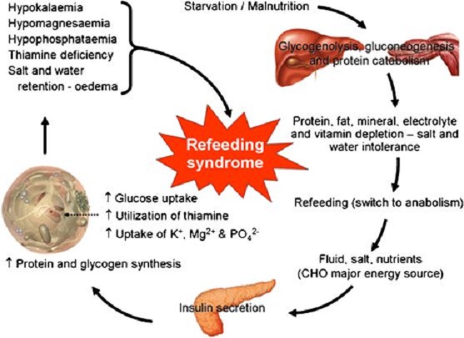
Refeeding syndrome is what happens when an individual’s long-standing state of catabolic metabolism is too rapidly treated with the necessary nutritional ingredients to restore the metabolic balance. Attempts to treat the chronic starvation of incarcerated victims in the Nazi -controlled camps after World War II resulted in death in many of them. It is a dangerous condition that occurs with reestablishment of adequate nutrition in malnourished and cachectic patients. More specifically, its occurrence has been reported during oral enteral and parenteral refeeding. I remember the case of a boy who was a “junk food junky”. He had just come down from a rope that he had been climbing in the gymnasium, illustrating his apparent physical fitness, but he then suddenly passed out. He was taken to a local hospital by ambulance when he was given glucose saline intravenously, a standard procedure. He had a series of bloodstained bowel movements and expired. We have known for years that introducing glucose intravenously to a long-standing thiamine deficient individual is dangerous. But how would this come to light in an emergency situation? To prevent the possibility of refeeding syndrome would demand the reason for this young man passing out be known in relation to his junk food diet.

Clinical features of refeeding syndrome are fluid-balance abnormalities, abnormal glucose metabolism and a deficiency of magnesium and potassium. In addition, thiamine deficiency dominates. Refeeding syndrome reflects the too rapid change from catabolic to anabolic metabolism, is often undiagnosed and some clinicians remain oblivious to its occurrence. Recognition reduces morbidity and mortality but there is no universal agreement as to its definition. A report exploring refeeding syndrome across seven cases found that each showed deficiencies and low plasma levels of potassium, phosphate, magnesium and thiamine combined with salt and water retention. It is interesting that salt and water retention are typical of thiamine deficiency. Similarly, a report exploring the incidence of Wernicke’s Encephalophy with anorexia nervosa found that 8 of 12 cases of anorexia investigated were afflicted with the full symptoms of the thiamine deficiency brain disease known as Wernicke encephalopathy. Anorexia affects 2.9 million people worldwide and is generally considered to be psychiatric loss of appetite. Both its active state and refeeding can be lethal.

Guidelines for refeeding patients with anorexia have been published by the European Society of Clinical Nutrition and Metabolism. Of 65 in-hospital patients studied, 14 were admitted more than once within the study period. Nine patients had minor complications in the first 10 days of replenishment. Four patients had transient pretibial edema (simple pressure with a finger below the knee resulted in dimpling). Three patients had what was described as organ dysfunction and two patients had severe hypokalemia (low potassium in the blood), all of which have been described as typical of thiamine deficiency. In fact, pretibial edema can be the only clinically obtainable evidence of thiamine deficiency. There is a high prevalence of thiamine deficiency in cancer patients. The prevalence of malnutrition is high in head-neck cancer patients, many of whom require artificial nutritional support or refeeding intervention. Refeeding syndrome is commonly encountered in the nutritional treatment of critical illness. However, guidelines and its occurrence in ICU patients remain unclear. Calorie restriction for several days and a gradual increase of its intake has been recommended. Thiamine deficiency brain disease is not too uncommon in parenteral nutrition. It has been reported that refeeding syndrome occurs in 4% of cases of parenteral nutrition, but failure of its recognition occurs in 50%.

Refeeding, Paradoxical Reactions, and Side Effects

Discerning readers of Hormones Matter have probably noticed that the subject of “paradox” has been mentioned a number of times and some time ago there was a comment that an article on the subject might be relevant. “Paradox” is a less severe form of RFS, a term that I have used to indicate that the patient’s expectation of improvement by nutritional replacement is often dashed because the symptoms become worse. Obviously, because pharmaceuticals are the usual and customary form of treatment, the worsening of symptoms immediately gives rise to the patient’s deduction that these are side effects. In a sense, they are indeed side effects, but the mechanism is very different from that caused by pharmaceuticals. The accentuation of symptoms represents a sudden switch from the prolonged state of catabolic/anabolic balance to that of anabolic/catabolic balance, whereas side effects of drugs are a direct effect of toxicity. This accentuation of symptoms seems to be directly related to the chronic nature of the malnutrition. It means that the unfortunate patient has been suffering for an extended period without the symptoms being recognized for what they represent. If the symptoms are correctly diagnosed at the outset of symptoms, the nutritional correction is easy and occurs rapidly. Paradox is because recognition comes after protracted malnutrition and is much more likely with intravenous nutritional correction. I always warned the patient before administration. However, the paradoxical worsening of symptoms may last as long as a month when vitamin therapy is used in oral administration. I have always told the patient that paradox is the best sign of ultimate improvement. For example, I was discussing the common symptoms of high calorie malnutrition with a nurse. She interrupted by telling me that I was describing the symptoms that she had been suffering for years. I suggested the nutrient replacement and she told me later that paradox had lasted for a good month but was then replaced with an absence of symptoms and an energy level that she had never previously experienced.

Modern Malnutrition

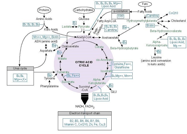
Readers must understand that chronic long-term malnutrition is common in America. However, it is not the same as the kind of malnutrition that is seen for example in Bangladesh, or that seen in advanced cancer, known as starvation. The kind of common malnutrition in America is because of an excess of calories and is often seen as an oxymoron. How can a high calorie diet possibly cause a potentially severe illness? The clinical expression of starvation is that of bodily attrition through catabolic breakdown leading to death, the kind of clinical situation underlined by Mother Teresa and caused by lack of any sort of food. People with high calorie malnutrition look entirely different and often constitute what I call the “walking sick”, because they are commonly seen as “problem patients” in the offices of physicians. They are often obese and their many complaints are most often diagnosed by physicians as “psychosomatic”. Their problem is too much food of the wrong sort. Thiamine/magnesium levels in the blood are usually perfectly normal if they are ever measured, giving rise to a physician’s refusal to diagnose the “absurd idea of a vitamin deficiency”. Thiamine activity is inside cells, so finding it in the liquid part (plasma) of the blood is meaningless. Like a “choked car engine” the non-caloric nutrients are overwhelmed by the excess of “empty” calories. It is energy production that is the core issue and is the reason for the multiplicity of symptoms. This is particularly true for deficiency of thiamine and magnesium because they are so essential to the processing of simple sugars. The indiscriminate ingestion of sweets has become a national calamity. Of course, thiamine and magnesium have to cooperate with many more non-caloric nutrients but their position in metabolism dominates the function of energy production.

So, high calorie malnutrition is an example of the effects of extremes, too much versus too little. The brain, heart and nervous system are the most affected organs because of their high energy requirement. The commonest symptom is fatigue but other common symptoms include “brain fog”, insomnia, a perpetual sense of anxiety or fear, heart palpitations, migraine, tension headaches, poor tolerance to heat and cold, unusual sweating particularly at night, diarrhea alternating with constipation, pins and needles in the extremities and vicarious body and limb pains. Because physicians in America have denied the possibility of vitamin deficiency disease, they usually interpret any abnormal laboratory studies to what they consider to be well-recognized and common diseases such as chronic fatigue syndrome. If lab studies are normal, then it is deemed to be “psychological”, a very unsatisfactory explanation to the patient.

When a person has been consuming simple carbohydrates in the form of sweets, including carbonated beverages and alcohol for a prolonged period, the general efficiency of cellular metabolism gradually declines. The three meals a day in some cases may be perfectly adequate but the dietary excesses may come for that person because of “the goodies” associated with social activities. Because the brain is most affected, the symptoms will be generated by the biochemical and electrical changes that follow. The symptoms are so variable that listing them all is virtually impossible. It has long been known that beriberi, the clinical expression of thiamine deficiency, had a long morbidity and a low mortality. The suffering experienced during its prolonged course was (and still is), however, an abysmal reflection of medical ignorance.

We Need Your Help

More people than ever are reading Hormones Matter, a testament to the need for independent voices in health and medicine. We are not funded and accept limited advertising. Unlike many health sites, we don’t force you to purchase a subscription. We believe health information should be open to all. If you read Hormones Matter, like it, please help support it. Contribute now.

Yes, I would like to support Hormones Matter.

Image: Nutrition in clinical practice—the refeeding syndrome: illustrative cases and guidelines for prevention and treatment.

This article was published originally on July 29, 2019. 

Refeeding Syndrome in the Context of Thiamine Deficiency

28687 views

Although this has been mentioned many times in posts and in the comments of readers on this website, there still seems to be a lack of understanding. The commonest complaints have been that “thiamine caused side effects” or “I was allergic to thiamine”, inevitably causing the complainant to discontinue it. I want to emphasize the important meaning of these seemingly adverse effects by illustrating a typical case in my own experience. First of all, please understand that thiamine deficiency has its major effects in the lower part of the brain. It is this part of the brain that controls the automatic (autonomic) nervous system that orchestrates the functions of all the organs in the body. Thiamine deficiency has its dominating effect by damaging this system and the result is known as dysautonomia.

Recognizing Thiamine Deficiency Syndromes

One day I was in conversation with a young woman and was trying to describe the huge number of symptoms that result from dysautonomia. When I finished listing them, I was surprised when she said that I had exactly described the symptoms that she had suffered for years. I had no prior knowledge of this, believing that she was completely healthy. She told me that this polysymptomatic condition had been present for as long as she could remember. Apparently it had never been understood by any physician that she had consulted and she had come to accept that it was “just the way that I am made”. She was in her early thirties and it must have required a lot of courage to do the work for which she was employed. Unrelenting fatigue dominated her life, and this is a major clue to her problem.

Symptom Exacerbation: Refeeding Syndrome

I advised her to start taking thiamine and magnesium supplements, starting with a low dose and advising her that the symptoms would become worse for an unpredictable period of time (refeeding syndrome). Note that this individual was known to be intelligent, was fully employed and that nobody was apparently aware that she had any health problems. Later she told me that after she started the supplements, for a month or more she had suffered an excruciating exaggeration of her many symptoms. Trusting that I knew what I was talking about, she persisted with the supplements. This is of great importance because without this information it might be interpreted as “side effects” and the nutrients withdrawn. It also would probably accompanied by anger and the ultimate symptomatic relief never experienced. Using her own words she then said “after about a month of taking the supplements, all my symptoms disappeared and my energy was better than any that I had experienced in my whole life”.

I will try to interpret what was happening here as an example of refeeding syndrome. It is important to understand that the many symptoms experienced by this woman were due to cellular energy deficiency in the brain. Their variability may have included emotional symptoms such as anxiety, depression, or anger without obvious cause because they would be the result of exaggerations of normal brain activity. The lower part of the brain is highly sensitive to energy deficiency and because it organizes all bodily functions, it can give rise to heart palpitations, chest pain, unusual sweating, pins and needles in the extremities, nausea, abdominal pain, vomiting, insomnia, constipation, diarrhea, or abnormal sense of balance including vertigo. Body pain that has no observable cause (hyperalgesia) or a pain response from a stimulus that does not usually cause pain (allodynia) may occur.

Refeeding Syndrome in Children

A 14-year old boy with sugar induced thiamine deficient eosinophilic esophagitis suffered agonies of hyperalgesia and screamed when I touched his abdomen (allodynia). Postural Orthostatic Tachycardia Syndrome (POTS) is quite a common variant which is particularly frightening to the patient. Let me emphasize once and for all, when symptoms like this go unrecognized, sometimes for years, they become temporarily exaggerated if the necessary nutrients are provided in too high a concentration. Whether this be a single vitamin, a group of vitamins or whole nutrition, this syndrome must be expected. A gradual introduction of the appropriate nutrients is mandatory. Because thiamine is so integral to energy metabolism, I found over the years that it was the most important. Because young children have not been exposed to malnutrition for too long because of their age, refeeding syndrome is seldom if ever encountered. The syndrome is directly related to the time of exposure to malnutrition and its severity. It is therefore an effect in adults and occasionally in adolescents..

Whether intelligence is a genetically determined gift or whether it is acquired during life, the brain consumes a disproportionate degree of energy that can only be met by an appropriate ingestion of food and water. If this is inadequate, symptoms begin to register the inadequacy by producing a sense of fatigue as the dominant one. It is the way that the brain signals its lack of cellular energy. The symptoms are easily removed if the underlying cause is recognized early. Because in many cases they are not recognized and the malnutrition may continue, it is not very surprising that cellular damage would be expected gradually to accrue. Perhaps chronic neurodegenerative disease may follow.

From Catabolic to Anabolic Metabolism

The normal states of damage and repair (anabolic metabolism) would be inadequate and a state of gradual breakdown and inadequate repair would be predicted (catabolic metabolism). Because thiamine deficiency causes the condition known as beriberi, I would like to state once more that the English translation of this Chinese word is “I can’t, I can’t”, severe, intractable fatigue being the dominating effect. Although the refeeding syndrome is poorly understood according to current medical literature it is apparently related to a rapid change from catabolic to anabolic metabolism. The misguided attempts to re-nourish the victims in concentration camps at the end of World War II resulted sometimes in their death. It is at least understood that correcting catabolic to anabolic metabolism, whatever produced the abnormal state, demands low doses of food in starvation and low doses of supplementary vitamins in the long term effects of high calorie malnutrition.

We Need Your Help

More people than ever are reading Hormones Matter, a testament to the need for independent voices in health and medicine. We are not funded and accept limited advertising. Unlike many health sites, we don’t force you to purchase a subscription. We believe health information should be open to all. If you read Hormones Matter, like it, please help support it. Contribute now.

Yes, I would like to support Hormones Matter.

This article was published originally on January 6, 2020. 

Thiamine Repletion, Calcium Management, and the Heart

17060 views

Individuals who have been chronically ill often show varying degrees of thiamine insufficiency ranging from functional deficiencies where, due to the energetic demands of an illness, diminished capacity of transporters and/or enzymes, and/or the high degree of oxidative stress, the need for thiamine outstrips the supply available via diet alone, to quite severe and frequently longstanding frank thiamine deficiency disease processes like beriberi or Wernicke’s encephalopathy. Depending upon the origins and chronicity of the deficiency, thiamine replenishment outside of hospitalization can be challenging and yet this is where it must take place, as physicians are reticent to consider or treat thiamine deficiency in office or hospital. Thiamine deficiency is neither recognized in the population nor accorded the resources required to resolve and as a result, patients are left to navigate the illnesses brought on by thiamine deficiency independently.

For some, thiamine supplementation is a simple process that nets noticeable improvements in symptomology almost immediately. For others, however, the process is grueling and marked by a period of time where symptoms become noticeably worse before improvement is seen. Replenishment of thiamine in longstanding deficiency, particularly when the heart is affected, is prone to a variety of ‘refeeding syndrome’ – type responses. Conventionally, refeeding syndromes involve disturbances of potassium, magnesium, and phosphate and develop when nutrients are re-introduced after a period of absence-based malnutrition, e.g. some form of starvation (see Figure 1.).

Figure 1. Refeeding syndrome.

refeeding syndrome
From: Nutrition in clinical practice—the refeeding syndrome: illustrative cases and guidelines for prevention and treatment

Symptomatically, these disturbances clearly affect heart function with troubling changes in rate, rhythm, and pressure. I suggest that the responses we see with more modern examples of chronic thiamine deficiency are generally caused by a higher calorie type of malnutrition; one where sufficient calories may be ingested absent sufficient nutrients. This is process is likely to be metabolically different than that of starvation or malnutrition. This means that while electrolyte disturbances often coincide, there may be other factors involved as well; factors that have yet to be fully understood.

Although the symptoms of refeeding noted in the high-calorie types of malnutrition may cause similar disturbances to heart function as the starvation-originated refeeding syndromes, given the distinct metabolic origins of these processes, different responses may be necessary. In some cases, the refeeding-like syndrome may be related to the formulation of thiamine used to treat the deficiency. There are several reasons for this, and our good friend Elliot Overton has begun to elucidate some of the chemistry involved. Across formulations, however, for a subset of patients thiamine repletion distinctly affects heart function even at micro-doses. Given that these patients are notably deficient in thiamine, that thiamine deficiency is absolutely requisite for mitochondrial function, for heart function, and for life itself, and its absence is directly associated with heart failure, how does one replenish thiamine when even the smallest dose sets off a cascade of troubling reactions?

With conventional treatment of wet beriberi, thiamine deficiency that affects the function of the heart, high doses of IV thiamine are provided for several days to a week in hospital, followed by high doses of oral thiamine for an extended period of time. The literature is rife with examples of success stories following this type of protocol. Absent from these case reports are instances of negative cardiac reactions to the high-dose thiamine. This suggests either these types of reactions do not occur by some function of the higher dosage, that they do occur but are mitigated by close consideration of electrolyte balance and/or other variables, or that they do occur but are simply not reported. Whatever the reasons, this would be useful information to practitioners and patients alike.

Assuming that these types of reactions occur, it is not clear why or by what mechanisms, beyond, of course, the electrolyte disturbances noted in refeeding of formerly starved individuals. This is important because while traditional interpretations of thiamine deficiency consider it rare and specific to a particular subset of patients for whom general nutrition and nutrient absorption is problematic (alcoholics, gastric bypass patients, parenteral feeding, anorexics, poverty-based malnutrition/starvation, pregnancy, with and without hyperemesis, and critical illness), it is not clear how those who develop beriberi in the presence of sufficient caloric intake, but with longstanding illness, differ metabolically from the traditional patient reported in the literature. And really, if we are honest with ourselves, we do not know much about the processes leading up to what are considered the more emergent cases of thiamine deficiency or even about the cases of heart failure where thiamine deficiency was likely a contributing factor but never considered. We have these pat, fully enshrined ideas about the ‘what and why’ of thiamine deficiency but rarely consider if or whether we know what we think we know.

With all of that in mind, I have been looking into potential mechanisms involved in the remodeling of heart tissue and function relative to longstanding thiamine insufficiency and the problems associated with thiamine treatment in these patients. I have observed that for some individuals, including those who have tested positive for thiamine deficiency and who clearly need thiamine, the addition of even the minutest amount of thiamine sets off a cascade of reactions that affect the pressure, rate, and rhythm of the heart. In these individuals, who may or may not have had previous, or at least previously recognized symptoms associated with cardiac function, the irregularities pose a difficult challenge for recovery; one for which the current literature has few direct answers.

I suspect these reactions involve an inability to smoothly switch from a previously hypoxic state to a marginally more normoxic one. I suspect also that the problem involves disturbed calcium (Ca2+) management adapted to the hypoxic environment that is then forced to re-equilibrate rapidly once respiration and oxidation kick on. Perhaps the response is akin to an ischemia/reperfusion process but one where longstanding molecular adaptations are involved versus an acute constriction of blood flow and oxygen. Regardless of the difference in time and scale, both the cause and the consequence involve altered Ca2+ flux, which is arrhythmogenic. If this is the case, it still begs the question about how we proceed with thiamine repletion, and perhaps also, how do we identify those most likely to respond negatively to thiamine? I do not have the answers to those questions yet, but perhaps if we unpack the relationship between mitochondrial function and calcium regulation we might find clues.

Mitochondrial Competence and Calcium Regulation

Calcium regulation depends upon functioning mitochondria and a steady stream of ATP. Mitochondrial functioning, in turn, depends upon the appropriate concentrations of Ca2+. The relationship between the two is reciprocal and dynamic. Failure in either leads to failure in both.

Mitochondrial nutrient deficiencies lead to reduced energy metabolism and utilization and ultimately molecular hypoxia. Since the heart requires an enormous amount of energy to maintain constant contractility, upwards of 15 times its weight in ATP – or about 6kg, every single day, mitochondria play a prominent role in heart function.

In the cases I see, presumed issues with Ca2+ mismanagement likely correspond to longstanding deficits in mitochondrial energetics evolving from some degree of metabolic inflexibility of the cardiomyocyte specifically, but likely, systemically. This metabolic inflexibility is both a consequence and a cause of insufficient thiamine, and likely other nutrients, that ultimately depress mitochondrial function and the capacity to produce ATP. The reduction in ATP then evokes a cascade of adaptive reactions, among them is a switch in fuel preference in the heart itself, from fatty acid oxidation to an increased reliance on glucose and anaerobic glycolysis; patterns consistent with systemic metabolic disruption. Additionally, the breakdown of amino acids is disrupted, leading to an accumulation of branched-chain amino acids, which further imperils energy production but also protein synthesis cascades. Finally, the decreased oxidative capacity that diminishes ATP output then continuously influences and is influenced by, Ca2+ dynamics creating a cycle of dysfunction. Arguably, it is these changes in mitochondrial metabolism that force alterations to cell and tissue morphology leading to the grossly observable pathology in later-stage heart failure.

Calcium Dynamics, Mitochondria, and the Heart: A Deeper Dive

Calcium dynamics in the heart are directly tied to the mitochondria at a number of junctions. Broadly, at physiological concentrations, Ca2+ activates oxidative phosphorylation (OXPHOS), e.g. ATP production. At supraphysiological concentrations, however, as we might see locally in the cardiomyocyte during the progression of heart disease to heart failure, Ca2+ inhibits OXPHOS. The inhibition of OXPHOS would presumably drive the shift in fuel source ultimately associated with heart failure. It would also drive calcium uptake, both by the mitochondria and sarcoplasmic reticulum (SR), altering cardiomyocyte contractility patterns. As mentioned previously, Ca2+ management by the mitochondria and SR are profoundly energy/ATP intensive processes.

Decrements in ATP availability, then lead to the poor uptake of Ca2+ by the mitochondria and SR, which then results in excess intracellular Ca2+ and impaired relaxation of the cell. This leads to high oxidative stress and increased reactive oxygen species (ROS) output; a pattern likely seen systemically. The high oxidative stress load caused by the overload of Ca2+ eventually stimulates mitochondrial permeability transition pore (mPTP) opening and mitochondrial swelling, resulting in mitochondrial injury, apoptosis, cardiac remodeling, and ultimately, the development of heart failure. Interestingly, a study in rodents found that with marginal thiamine insufficiency, the ensuing accumulation of pyruvate opened the mPTP but in a low-conductance state that apparently does not lead to cell death. The authors postulate that this may be a protective response that tempers the excess pyruvate and conditions the heart to reduced oxygenation.

Among the mechanisms by which Ca2+ regulates OXPHOS, all are nutrient-dependent; nutrients that are likely insufficient in metabolic disease and the progression to heart failure (see Figure 2.).

Figure 2. Mitochondrial nutrients.

Mitochondrial nutrients
From: Thiamine Deficiency Disease, Dysautonomia and High Calorie Malnutrition.

Moreover, each of these enzymes is activated appropriately only in the presence of the requisite nutrients and at low levels of Ca2+. At a higher concentration of Ca2+ and/or in the absence of the other nutrient cofactors, Ca2+ inhibits these enzymes, effectively shunting ATP production.

  • Calcium activates the pyruvate dehydrogenase complex (PDC) both directly by binding to pyruvate dehydrogenase, or indirectly by upregulating inactivation processes and binding to pyruvate phosphatases. The PDC is exquisitely sensitive to thiamine and magnesium.
  • Calcium stimulates the alpha-ketoglutaric dehydrogenase enzyme complex (KGDH), a rate-limiting enzyme within the Krebs cycle, but only at low concentrations and in the presence of magnesium and thiamine. At high concentrations and in the absence of sufficient Mg2+ or thiamine, Ca2+ inactivates KGDH leading to impaired energy metabolism and poor calcium management in the mitochondria.
  • Calcium upregulates mitochondrial complex I, III, IV, and the Vmax of complex V, leading to KGDH inhibition when appropriately operational, and once again, likely only in the presence of the requisite nutrient cofactors (see Figure 2).
  • Each of the processes above is energy dependent, necessitating sufficient ATP, but also, use the energy released by these reactions to reduce the electron the carriers nicotinamide adenine dinucleotide (NAD+) to NADH and Flavin adenine dinucleotide (FAD+) to FADH2. Both NAD and FAD require niacin and riboflavin, respectively. Calcium activates FAD-glycerol phosphate dehydrogenase and NAD-isocitrate dehydrogenase, but again, only at low concentrations and in presence of sufficient ATP and magnesium.

In sum, the relationship between the mitochondria and Ca2+ regulation is largely dependent upon the presence of critical micronutrient cofactors. Insufficiencies imperil ATP production and Ca2+ management, which then leads to a cycle of dysfunction likely at play in heart failure.

Calcium Capture and Release by the Sarcoplasmic Reticulum

If we move out of the mitochondria into the cytoplasm, we see similar sequences of events play out in the SR. The SR within the cardiomyocyte bears the brunt of responsibility for Ca2+ management in normally functioning hearts. Indeed, some research suggests that it is only when things go awry that the mitochondria step in to become the primary Ca2+ sinks, sequestering the excess. Otherwise, mitochondrial Ca2+ stores reflect those of the cytosol due to the constant influx and efflux of the cation via a number of transport channels, uniporters, and exchangers.

Here again, Mg2+ is involved. Magnesium, it appears, competes with Ca2+ at the ryanodine receptor, a critical Ca2+ receptor in the SR, to inhibit the release of intracellular Ca2+, and essentially temper Ca2+ activation/release. As with the mitochondria, SR activation is possible only at low concentrations of Ca2+ and when Mg+ and ATP are complexed together. High concentrations of Ca2+ inactivate the receptor, inhibiting the further release of Ca2+ into the cell. Similarly, high concentrations of Mg2+ also inhibit the receptor. All of this has to be exquisitely timed and balanced. Too much or too little, too soon or too late induces asynchrony in the contractility of the heart muscle and is one of the variables in the progression to heart failure.

Of note, caffeine increases the sensitivity of the ryanodine receptor by ~20-50 times. This means that less Ca2+ is required to open the receptor and release Ca2+ stores, potentially leading to high intracellular Ca2+ levels. Coffee and tea, the two more popular carriers of caffeine, are also high in thiaminase, a thiamine breakdown enzyme. Given how many of us are addicted to coffee and the caffeine it holds, this is problematic for both thiamine maintenance and heart function.

Problems with Thiamine Repletion

With all of this, I still have no idea what is causing the negative reaction to thiamine repletion in some patients. I do have a few ideas though, but none are fully worked out just yet. My gut tells me it is a matter of too much too soon and an inability to smoothly switch from the previously hypoxic state that relied on anaerobic glycolysis for ATP to a more normoxic state and an upregulation of OXPHOS. To that end, I suspect that the problem rests with three variables, the lack of substrate fuels for the newly upregulated OXPHOS (glucose and fatty acids), a lack of additional micronutrients (riboflavin, niacin, and magnesium, but possibly also Coq10) and an inability to manage the increase in carbon dioxide resulting from the increased oxygen utilization. All of this, of course, would impact Ca2+ homeostasis. I suspect the key roadblock in the process is alpha-ketoglutaric dehydrogenase, both as a rate-limiting complex in ATP production, ROS regulation, and Ca2+ regulation, but also, I suspect it is somehow tied to the regulation of the carbonic anhydrase enzymes that manage blood gas exchange. How though, I am not sure.

First possibility: Thiamine kick starts previously quiescent enzymes increasing ATP production via OXPHOS and Ca2+ induced excitation begins. Rather than appropriately sequester excess Ca2+, the longstanding reduction of Ca2+ in the cell is suddenly upregulated by SR receptor open time. That then feedbacks and shuts off the alpha-ketoglutaric dehydrogenase complex, which would again reduce ATP, while simultaneously increasing ROS. Alpha keto glutarate is a primary anti-oxidant next to glutathione. Elevated ROS and mitochondrial Ca2+ underpin several variables in heart failure including hypertrophy, fibrosis, and contractile dysfunction. If thiamine is given with insufficient magnesium and the other nutrients required for proper mitochondrial functioning (Figure 2.), alpha-ketoglutaric dehydrogenase would become inhibited or asynchronous to the relative increase in thiamine and Ca2+.

Second possibility: the increased thiamine fails to initiate OXPHOS sufficiently and continues to feed the anaerobic cycle shunting more pyruvate to lactate dehydrogenase with an ensuing buildup of lactate and a yet insufficient amount of ATP. With longstanding thiamine deficiency and metabolic dysfunction, the fuel preference of the heart adapts and switches from a primarily fatty acid-derived form of oxidation (60-70% fatty acid preference over glucose) to one that is entirely dependent upon large amounts of glucose via anaerobic fermentation. The shift back would be difficult, especially if thiamine is given quickly and in large amounts, or possibly any amount, absent the other nutrient co-factors. While thiamine is critical in the first steps of fatty acid metabolism, in alpha oxidation, to get through beta-oxidation, one needs carnitine, and vitamins, B2, B3, and B5. Absent those co-factors, and importantly, absent the substrate fatty acids to metabolize, we might yet get held up. Conceivably, this might also happen absent sufficient glucose to sustain the long-entrenched baseline production of ATP via anaerobic metabolism while OXPHOS is upregulating. While glucose is typically contraindicated in the thiamine repletion, as it is often a causative factor in the shifted metabolism in the first place, it is possible that in some instances it may be warranted. If the heart’s preferred substrate is not available then no matter how much we upregulate the OXPHOS pathway, energy production would falter.

Third possibility: Here, I have not yet fully put the pieces together, but briefly, it is likely the longstanding thiamine deficiency changes the regulation of blood gases and that this shift is problematic when oxygen status changes. We know that thiamine deficiency stabilizes the hypoxia-inducible factor (HIF) proteins. HIF proteins are the key regulators of the hypoxia cascades. That means, the absence of thiamine causes a sort of molecular hypoxia, an inability to utilize oxygen for the production of energy and to traffic it appropriately in the hemoglobin >oxyhemoglobin. In the case of longstanding thiamine insufficiency or deficiency, we have all of these underlying adaptations that have come into play to compensate, including those involved in the management and usage of oxygen. Since thiamine modulates this process and has been noted to be a carbonic anhydrase inhibitor both deficiency and excess would be problematic. I suspect that when we shift one side of the equation e.g. add thiamine back to the process, effectively upregulating the use of O2 and the production of ATP, the compensatory reactions of the other side, the re-regulation of Ca2+ and the carbonic anhydrase (CA) enzymes, falters. The CA  enzymes are responsible for balancing the consumption of O2 with the production of carbon dioxide (CO2). Asynchrony here would be problematic. This in turn would stress mitochondria and evoke a whole host of potentially negative reactions. Among them, I think alpha-ketoglutaric dehydrogenase downregulation is a part, but I have no evidence of this yet.

That is what I have thus far. As I continue to put pieces together, I will publish subsequent posts. For now, though, I think the key takeaway is that when replenishing thiamine, we have to be cognizant of the other macro and micronutrients involved in the production of energy, except in acute, immediately life-threatening cases, proceed thoughtfully. What that means clinically, and to those individuals for whom even the slightest amount of thiamine sets off deleterious reactions in the rate, rhythm, or pressure of the heart, I do not yet know. Perhaps others will chime in with theories.

We Need Your Help

More people than ever are reading Hormones Matter, a testament to the need for independent voices in health and medicine. We are not funded and accept limited advertising. Unlike many health sites, we don’t force you to purchase a subscription. We believe health information should be open to all. If you read Hormones Matter, and like it, please help support it. Contribute now.

Yes, I would like to support Hormones Matter. 

Feature image by Gerd Altmann from Pixabay.