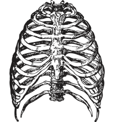
Many years ago, I wrote a short post about some unique research linking manganese deficiency in deer antlers to osteoporosis in humans. I had long since forgotten about it. Recently, someone left a comment about a connection between manganese and thiamine. It turns out that manganese deficiency diminishes tissue storage and synthesis of thiamine. So, in addition to potentially driving osteoporosis, manganese is influences thiamine availability. Given these connections, I thought I might revisit the research.
Deer Antlers, Manganese, and Osteoporosis
The dominant theory about osteoporosis in humans and animals alike, is that deficiencies in calcium and/or vitamin D drive osteoporosis. Although both impact bone density, in many circumstances, it is not clear whether these factors are causative, coincidental, or a result of some other variable. There is a growing body of evidence suggesting that while calcium and vitamin D deficiencies are problematic and can indeed induce problems with bone development and strength, another nutrient may also be involved: manganese.
Manganese, a trace metal found naturally in a wide variety of foods, including nuts, plants, and shellfish, is a cofactor for many enzymes. Deficiency is considered rare, but it is infrequently measured. Much of the research on manganese focuses on its toxicological properties when exposed to excessive amounts, usually occupationally. Since manganese accumulates in bone, excessive manganese is just as problematic for bone health as deficiency. That said, manganese is integral for normal processes of bone development and integrity. A great review can be found here.
Returning to the deer antler paper for moment, the authors posited a mechanism that I had not yet seen fully explored – that declining bone calcium observed with osteoporosis is linked to low manganese. Specifically, they argued that manganese deficiency impairs calcium uptake and integration in the bone and thus drives osteoporosis. This is claim has been repeated frequently since the work was published in 2010 but it is rarely discussed more thoroughly, at least that I could find. The actions attributed manganese in bone development include: enzyme cofactor activity; protection of osteoclasts from oxidation processes during growth and remodeling via the mitochondrial antioxidant MnSOD (manganese superoxide dismutase); cartilage and collagen synthesis; and its ability to stimulate other signaling pathways involved in bone development and integrity. Certainly, all of this is important, but I find the role it may play in calcium management is particularly interesting.
In the deer antler study, researchers found that during an especially cold winter manganese in the plant matter consumed by the deer declined. In parallel, manganese but not calcium concentrations in the antlers declined, as did antler tensile strength and bone integrity, while fractures increased. Since during development about 20% of circulating calcium is shuttled preferentially to the antlers, stable calcium concentrations in the antlers made sense. When the antlers of wild deer were compared to fed deer who survived that winter, antlers of the fed deer showed no declines in manganese or in antler integrity. This begs two questions, how does manganese influence bone calcium and is there a similar relationship between low manganese and increased osteoporosis in humans?
Regarding the first question, the research on mitochondrial manganese/calcium in bone is limited. That said, there is a fair amount of research on other tissues. A study from the 1980s showed that manganese altered the flux of calcium into and out of the rodent liver, heart, and brain mitochondria favoring influx. In other words, when manganese was present at concentrations consistent with those that might exist in real life, the mitochondria pulled in more calcium than they spit out. When magnesium was present simultaneously, the reaction was larger. More recent studies show that when manganese is present at supraphysiological concentrations, it overrides calcium influx, slows efflux, and more manganese is pulled into the mitochondria than calcium. Here, the mitochondria appear to buffer excessive manganese over calcium, but in doing so, elicit changes in mitochondrial and cellular calcium kinetics potentially leading to excitotoxic cell death – which we will discuss momentarily.
Regarding the second question, human bone contains ~40% of total body manganese and so it is possible that deficiency could lead to similar decrements in bone integrity. Several studies published since the deer antler study have linked low manganese to poor bone health and manganese supplementation to improved bone health. Interestingly, individuals with neurodegenerative disorders like Parkinson’s, Alzheimer’s, dementia, etc., show a higher propensity for osteoporosis compared to elderly patients of same age without neurodegenerative disorders. Obviously, there is a lot going on with these conditions, but I cannot help but wonder if when dietary manganese is low, bone manganese is consumed or released to favor brain manganese, thus leading to osteoporosis first, and neurodegeneration secondarily when the deficiency is prolonged. If that is the case, then osteoporosis in the elderly may be an early indicator of not only low manganese but impending brain dysfunction. This is speculative, of course, but I think a case can be made, especially when we consider the role of manganese in thiamine storage and thiamine’s role in calcium trafficking and management.
Thiamine and Calcium
Thiamine or vitamin B1, is integral to mitochondrial health. Mitochondrial health is integral to calcium management within the cell. The entry of calcium into the cell is excitatory. It tells the cell to do something; to contract a muscle, fire a neuron, migrate somewhere, grow and differentiate, etc.. This process is tightly regulated, as too much calcium or too much too fast, can cause damage or even death to the cell. This is called calcium-induced excitotoxicity.
There are multiple mechanisms to protect against this, but the primary mechanism is calcium sequestration by the mitochondria and interaction with another set of organelles called the endoplasmic reticula (ER). The mitochondria within the cell effectively mop up excess calcium, store it and then release at a more appropriate time and at a rate that is more manageable for the cell. This of course, happens only if the mitochondria have sufficient resources to function properly. What are those resources? Nutrients.
I won’t bore you with details, as I have written hundreds of articles on this topic (search here), but briefly vitamins and minerals are required for the mitochondria to perform their requisite tasks and deficiencies limit or block their activity. The tasks mitochondria are responsible for include: making energy/ATP, synthesizing other proteins involved in cell function and development, managing redox and detox processes, life/death cycles of the mitochondria but also of the cell itself, and calcium management. Thiamine is a key nutrient involved in all of these processes but is absolutely critical to the production of ATP. ATP is the energy or fuel that cells use to do all of things they need to do. What this means is that low thiamine leads to insufficient ATP, which in turn leads to poor cell function, and eventually, cell damage and death. Oh, and ultimately, our death too. We cannot even breathe without sufficient ATP. So, there is that too.
Back to thiamine and calcium. Again, I have written about this as well (here, here) as it relates to the heart, but this process is conserved across different cell populations. Briefly however, insufficient thiamine causes mitochondrial mismanagement of calcium. This, I believe happens in stages. In the early stages of deficiency, mitochondria will ramp up and store any excessive calcium to temper signals of excitation. This is a protective measure that essentially downregulates cell activity. Even so, it is an energy intensive process and so as energy wanes, so too does the ability to store and manage calcium effectively. Disordered autonomic function, also known as dysautonomia, is an indication of inadequate thiamine, which in turn means poor calcium management and disordered neural signaling in the brainstem and cerebellum (and elsewhere, but I digress).
When thiamine deficiency becomes more pronounced, either by chronicity or with a severe or overwhelming stressor, mitochondrial tempering fails and previously stored calcium is released too quickly. This causes a rapid efflux of calcium into the cell, a calcium tsunami of sorts that can be considered an internal excitotoxic event. Remember, excitotoxicity leads to cell death.
Manganese and Thiamine
While all of this is cool, the question remains, how does manganese effect thiamine? Based upon studies done many decades ago using rodents, researchers found that manganese increases the storage in multiple tissues and the synthesis of thiamine into its more active form, thiamine pyrophosphate (TPP), also called thiamine diphosphate (ThDP/TDP). A study from 1954 found that the addition of manganese to the diet increased liver thiamine and intestinal thiamine significantly. Interestingly, with low manganese, storage of thiamine in the small intestine is about 1/3-2/3 that of the liver, but with manganese, it exceeds liver storage. (Another digression: could this be a mechanism of SIBO? There is already evidence of low thiamine in SIBO, maybe low manganese is involved as well?)
A Russian study in 1985 found that total and bound thiamine content (TPP) increased in the blood, liver, heart and brain tissue while free thiamine decreased:
Addition of manganese to the diets promotes an increase of the total thiamine content in the blood and the liver, heart and brain tissues. This trace element appreciably changes the correlation between different thiamine fractions. The free vitamin B1 level in the blood and tissues decreases, while the level of its bound form (pyrophosphatic) increases. All the administered manganese doses induced a statistically significant reduction of pyruvic acid concentration in the blood.
I could not access the full study, but from the abstract it is important to note that manganese appears to increase TPP – the activated component of thiamine, which then reduces pyruvic acid content in the blood. High pyruvic acid is a key indicator of insufficient thiamine.
Other studies found similar relationships (here, here), but these relationships were not straightforward and likely to involve the interaction between other minerals and metals. Additionally, I was not able to find research untangling the thiamine>manganese relationship in humans. That said, I did find one study linking manganese to the activity of thiamine pyrophosphokinase, the enzyme responsible for phosphorylating/activating free thiamine and turning it into thiamine pyrophosphate/TPP. Granted the study was conducted in 1975, in a parsley leaf, and so, translating it to humans may be a leap, but if the human enzyme can utilize manganese, this would directly link manganese to thiamine. I should note that other enzymes accept multiple divalent cations. Pyruvate kinase (PK), for example, can substitute manganese for magnesium. The kinetic properties are different between a manganese activated versus magnesium activated PK.
Another potential mechanism at this same enzyme is via the binding to the ATP molecule. Pyrophosphokinase activity is dependent upon magnesium bound to ATP (MgATP). Manganese and magnesium share binding sites and may be somewhat interchangeable on several thiamine dependent or adjacent enzymes including a number of cytosolic and mitochondrial enzymes involved in bioenergetics. The determining kinetics of when an enzyme binds with manganese versus magnesium and to what end is unclear, at least to me, and at this point in time and so all of this is just speculation. Despite needing more research, I believe that manganese and thiamine are important components of bone integrity and when deficient create problems via the pattern below.
Low manganese →low TPP→ altered cellular calcium handling → impaired bone metabolism → osteoporosis
Final Thoughts
Barring the 1975 study mentioned above and few other studies by that same group, there seems to be very little research on the interactions between thiamine and manganese at the enzyme level. Similarly, aside from the few studies conducted in early part of the 20th century through about 1985 on how manganese affects thiamine concentrations in rodents, not much else has been done. All work since seems to have focused exclusively magnesium. Obviously, magnesium is important, however, if manganese and magnesium bind interchangeably on a variety of enzymes, including those that influence thiamine activity, it would seem to me that these are things we ought to be exploring too. I would venture that there is a whole compendium of important interactions we have missed that influence thiamine activity, and thus, mitochondrial capacity.
We Need Your Help
More people than ever are reading Hormones Matter, a testament to the need for independent voices in health and medicine. We are not funded and accept limited advertising. Unlike many health sites, we don’t force you to purchase a subscription. We believe health information should be open to all. If you read Hormones Matter, like it, please help support it. Contribute now.
Yes, I would like to support Hormones Matter.
Photo by Markus Winkler on Unsplash


I’ve found a clear relationship for me between manganese and thiamine.
I developed intense right-sided sciatica: deep pain in my right buttock, a sharp “zinging” sensation in my right tibialis anterior that caused my lower leg to jump, and episodes where my right leg would give out. Around the same time, my lower back and sacrum began popping when I turned over at night.
My chiropractor prescribed Standard Process Ligaplex (manganese). The recommended dose was quite high. At that dose, my central sleep apnea (CSA) returned with a vengeance. CSA is my personal red-flag symptom that I’ve crossed “below the black line” into thiamine deficiency.
I had to drastically reduce the manganese and triple my thiamine dose. I continued Ligaplex at just one tablet per day (55 mg), and—unexpectedly—my sciatica resolved. I’m not sure why manganese helped, as that wasn’t the original goal, but I was extremely grateful.
Since then, the sciatica has returned several times, and each time it has resolved again after taking about 25 mg of manganese daily for a couple of weeks.
I later found a study suggesting an interaction between thiamine and manganese, though it was beyond my basic biochemistry understanding. What mattered was knowing the connection exists: whenever I take extra manganese, I must double or triple my thiamine (thiamine mononitrate and allithiamine) to avoid deficiency symptoms.
I’ve noticed a similar pattern with copper. After reading the April 2, 2025 HormonesMatter article on copper, I added a multi-mineral containing 1 mg of copper. I initially felt more energy, but my central sleep apnea flared again. In this case, tripling thiamine wasn’t enough—I had to cut the copper dose in half (to 0.5 mg) while keeping thiamine doubled.
If I could start my thiamine journey over, I would have taken a multimineral from the beginning, alongside thiamine, magnesium, and riboflavin.
I also want to thank AMZ for discussing di-calcium phosphate—it appears to give me more energy as well.
The consistent pattern for me is this: correcting mineral deficiencies rapidly increases thiamine demand. If I don’t increase thiamine accordingly, there isn’t enough left to keep me breathing normally at night.
If anyone knows of a doctor or health coach in the Tucson area who truly understands thiamine and mineral biochemistry, I would greatly appreciate a referral.
Hello. What is known about HPV vaccine injury to date?
I found various articles on this website but they are old, has new knowledge been acquired since then?
I also saw on most of those articles you were making a survey asking for patients to share their symptoms. Were the results of this surveys ever published?
Sadly, the research was never completed and we had no funding to continue. I haven’t had any writers in a while and so, while there is likely a lot more research on the HPV vaccine, I haven’t been able to cover it.
I was injured by the HPV vaccine in 2024 and since then I have been suffering everyday.
I am tired and very sad, I don’t know what to do.
I am so sorry. Would like to share your story as a post and perhaps we can generate some interest and insight regarding how to help you heal?
Sounds like a good idea but since I haven’t found any relief I don’t the point on sharing my story now.
Did you ever figured out why the HPV vaccine causes the issues it causes?
The point is to show other women the risks. It’s also about documenting what you experienced so that perhaps someone out there can you help find something that helps you to heal. As far as what about the HPV vaccine in particular is so problematic – there is a long list of problems, but the reality is that so many of us are one medication or vaccine away from serious illness that it is difficult to dissociate the effects of one vax or one med from the laundry list of all of the others that we are exposed to and that cause horrible adverse reactions. The more you dig into the mechanisms of these drugs/vaxes, it becomes abundantly clear that most are no more than poisons. Regarding how to heal, I am big proponent of working from the mitochondria up and out, which means nutrients, and ultimately thiamine. Read up on mitochondria and thiamine here on the site and see if any resonates. Please consider sharing your experience too.
Do you ever figured what was wrong with the HPV vaccine victims? Based on your observations, what was causing the issues on them?
For many, it involves thiamine deficiency. Read up on thiamine on the site.
Yes, I’ve read a lot of the stories here. I was just wondering if that was still the current consensus.
It as never been the consensus, conventional medicine does not consider thiamine an issue, but it is the reality. Deficiency is more common than recognized and the breadth symptoms is broad. Here is an overview article. https://pmc.ncbi.nlm.nih.gov/articles/PMC8533683/